1,267 posters, 47 videos, 13 topics, 4 sessions, 853 authors
ePostersLive by SciGen Technologies S.A. All rights reserved.
SOHO 2026 Annual Meeting
September 9 - 12, 2026 | George R. Brown Convention Center, Houston, Texas

MM - 1322
Clinical Landscape and Prognostic Impact of Extramedullary Disease in Multiple Myeloma: A Large Single-Center Retrospective Analysis
Poster Presenter
Authors
Part of Topic
Multiple Myeloma (MM)
Clinical Landscape and Prognostic Impact of Extramedullary Disease in Multiple Myeloma: A Large Single-Center Retrospective Analysis
Nesrine Ben Sayed1 , Ameni Ben Amor1 , Amira Rahal1 , Wafa Chanbeh1 , Nadia Sassi1 , Haifa Regaieg1 , Yosra Ben Youssef1
1 Farhat Hached, Sousse, Tunisia
Abstract
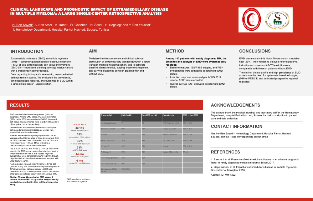
Background: Extramedullary disease (EMD) in multiple myeloma (MM)—comprising paramedullary osseous extension (PMD) or true extramedullary soft-tissue involvement (EMD-S)—represents a biologically aggressive variant with a traditionally poor prognosis. Data regarding its impact in real-world, resource-limited settings remain sparse. We evaluated the prevalence, clinicopathologic features, and outcomes of EMD within a large single-center Tunisian cohort.
Methods : Among 140 patients with newly diagnosed MM, the presence and subtype of EMD were systematically recorded. Baseline features, ISS/R-ISS staging, FISH cytogenetics, induction response (IMWG 2014 criteria), ASCT rates, and OS were analyzed according to EMD status.
Results : EMD was identified in 40/140 patients (29%) at diagnosis. Among EMD cases, PMD predominated (55%), while 35% presented with EMD-S. Bone and soft-tissue plasmacytomas were found in 24% and 3% of the global cohort, respectively. Involved sites included complex vertebroparaspinal, pelvic, and maxillofacial masses, as well as rare visceral/retroperitoneal masses. Patients with EMD were younger (median, 57 vs 59 years) and exhibited higher rates of bone involvement (98% vs 75%) but lower rates of anemia (38% vs 71%) and renal impairment (10% vs 41%), compared with patients without EMD, reflecting a predominantly osseous disease burden. ISS 3 (42% vs 57%) and R-ISS 3 (35% vs 45%) scores were lower in the EMD group, suggesting standard staging may underestimate risk in this subset. High-risk cytogenetics were comparable (22% vs 26%), although highrisk clinical classification was more frequent in EMD (80% vs 72%). Postinduction, rates of ≥VGPR (58% EMD vs 54% non-EMD), CR (22% vs 21%), and primary refractory disease (16% vs 17%) were similar between groups. ASCT was performed in 33% of EMD-group patients versus 26% in non-EMD–group patients. Relapse occurred in 53% versus 61% of patients. Median OS was 44 months for EMD versus 6 months for non-EMD, a paradox likely driven by survival data availability bias in this retrospective study.
Conclusion : EMD prevalence in this North African cohort is notably high (29%), likely reflecting delayed referral patterns. While induction response and ASCT feasibility were comparable with those of patients without EMD, the distinct clinical profile and high prevalence of EMD underscore the crucial need for systematic baseline imaging (MRI or PET/CT) and dedicated prospective regional registries.
ASCT: autologous stem cell transplantation, CR: complete response, FISH: fluorescence in situ hybridization, IMWG: International Myeloma Working Group, ISS: International Staging System, MRI: magnetic resonance imaging, OS: overall survival, PET/CT: positron emission tomography/computed tomography, R-ISS: Revised International Staging System, VGPR: very good partial response
Keywords MM, extramedullary disease, osseous extension, soft-tissue, outcomes, real-world, resource-limited setting