875 posters, 25 topics, 3,440 authors, 1,061 institutions
ePostersLive by SciGen Technologies S.A. All rights reserved.
SAGES 2026 Annual Meeting Emerging Technology Session
March 25-28, 2026 | Tampa, FL, USA

P578
Central Tendon Diaphragmatic Hernia: Surgical Management of a Paraesophageal Look-Alike
Poster Presenter
Authors
Affiliations
Part of Topic
Foregut
Introduction
•Diaphragmatic hernias located near but distinct from the esophageal hiatus are rare and often misidentified as paraesophageal hernias.
•CT is often (but not always) capable of clarifying the location of the diaphragmatic defect.
•Localization of the defect is important for preoperative diagnosis and operative planning.
Case Presentation
•A 67-year-old female was referred for evaluation of a large paraesophageal hernia detected on chest radiograph during a cough workup at an outside facility.
•Symptoms include intermittent heartburn, postprandial upper abdominal pain, bloating, and rare regurgitation.
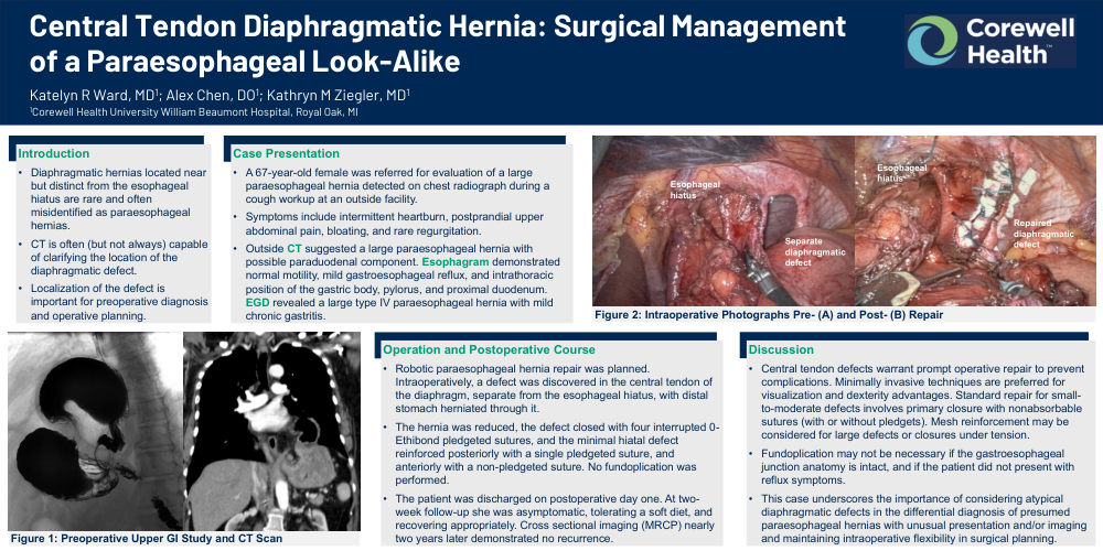
•Outside CT suggested a large paraesophageal hernia with possible paraduodenal component. Esophagram demonstrated normal motility, mild gastroesophageal reflux, and intrathoracic position of the gastric body, pylorus, and proximal duodenum. EGD revealed a large type IV paraesophageal hernia with mild chronic gastritis.
Operation and Postoperative Course
•Robotic paraesophageal hernia repair was planned. Intraoperatively, a defect was discovered in the central tendon of the diaphragm, separate from the esophageal hiatus, with distal stomach herniated through it.
•The hernia was reduced, the defect closed with four interrupted 0-Ethibond pledgeted sutures, and the minimal hiatal defect reinforced posteriorly with a single pledgeted suture, and anteriorly with a non-pledgeted suture. No fundoplication was performed.
•The patient was discharged on postoperative day one. At two-week follow-up she was asymptomatic, tolerating a soft diet, and recovering appropriately. Cross sectional imaging (MRCP) nearly two years later demonstrated no recurrence.
Discussion
•Central tendon defects warrant prompt operative repair to prevent complications. Minimally invasive techniques are preferred for visualization and dexterity advantages. Standard repair for small-to-moderate defects involves primary closure with nonabsorbable sutures (with or without pledgets). Mesh reinforcement may be considered for large defects or closures under tension.
•Fundoplication may not be necessary if the gastroesophageal junction anatomy is intact, and if the patient did not present with reflux symptoms.
•This case underscores the importance of considering atypical diaphragmatic defects in the differential diagnosis of presumed paraesophageal hernias with unusual presentation and/or imaging and maintaining intraoperative flexibility in surgical planning.
Figure 1: Preoperative Upper GI Study and CT Scan
Figure 2: Intraoperative Photographs Pre- (A) and Post- (B) Repair