875 posters, 25 topics, 3,440 authors, 1,061 institutions
ePostersLive by SciGen Technologies S.A. All rights reserved.
SAGES 2026 Annual Meeting Emerging Technology Session
March 25-28, 2026 | Tampa, FL, USA

P571
Delayed Presentation of Boerhaave Syndrome after Renal Transplantation
Poster Presenter
Part of Topic
Foregut
Introduction:
Boerhaave syndrome is a rare, spontaneous esophageal perforation affecting 3.1 per 1 million people annually. Mortality rates are up to 25% when treatment is initiated within 24 hours, and up to 60% after 48 hours, underscoring the importance of early diagnosis and management. Due to immunosuppression, transplant patients may have atypical presentations, and are especially susceptible to infectious complications. Here, we describe a renal transplant patient with delayed presentation and diagnosis of Boerhaave syndrome.
Case Description:
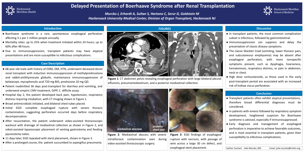
A 68-year-old male with history of end-stage renal disease, diabetes, and hypertension, underwent deceased donor renal transplant. Induction immunosuppression included rabbit-antithymocyte globulin with methylprednisolone. His maintenance regimen at discharge was belatacept, mycophenolic acid 720 mg twice daily, prednisone 10mg daily. He was readmitted 36 days after transplant with diarrhea and vomiting. Empiric treatment for cytomegalovirus was initiated while awaiting results of viral titers, gastrointestinal pathogen panel, and clostridium difficile toxin assay. Hospital day 2, he developed back pain, hypotension, and respiratory distress, ultimately requiring intubation. Computed tomography scan showed esophageal perforation with large bilateral pleural effusions, pneumomediastinum and a posterior mediastinal collection. Broad spectrum antimicrobials were initiated and bilateral chest tubes placed. Initial esophagoscopy revealed complete esophageal rupture with severe thoracic contamination, suggesting that perforation occurred days before respiratory decompensation. After resuscitation, the patient underwent video-assisted thoracoscopic washout with drainage of mediastinal collection, and robot-assisted laparoscopic placement of venting gastrostomy and feeding jejunostomy tubes. Eleven days later, an esophageal stent was placed, traversing a 30 centimeter defect. After a prolonged course, the patient ultimately succumbed to aspergillus pneumonia.
Discussion:
In transplant patients, the most common subset of complications are infectious, followed by gastrointestinal. Immunosuppression can suppress and delay the presentation of classic disease symptoms. The classic Mackler triad (vomiting, lower thoracic pain, subcutaneous emphysema) may be absent after esophageal perforation, with more nonspecific symptoms present, including dysphagia, hoarseness, hematemesis, respiratory distress, or pain in abdomen, neck or chest. Additionally, high dose corticosteroids, as used in the early post-transplant period, are associated with an increased risk of hollow viscus perforation.
Conclusion:
Transplant patients often exhibit atypical presentations, therefore a broad differential diagnosis should be considered. In patients with emesis followed by respiratory symptom development, heightened suspicion for Boerhaave syndrome is advised, especially if immunosuppressed. Early diagnosis and management of esophageal perforation is imperative to achieve favorable outcomes, and is most essential in transplant patients, who are especially susceptible to secondary infectious complications after perforation.