875 posters, 25 topics, 3,440 authors, 1,061 institutions
ePostersLive by SciGen Technologies S.A. All rights reserved.
SAGES 2026 Annual Meeting Emerging Technology Session
March 25-28, 2026 | Tampa, FL, USA

P138
Bleeding Lateral Oblique Hematoma in a Coagulopathic Female with Hemophilia B
Poster Presenter
Part of Topic
Acute Care
Bleeding Lateral Oblique Hematoma in a Coagulopathic Female with Hemophilia B
Valerie Fiore, DO1, Jovia Williams2, Marc Knezevic-Maragh, MPH2, Caleb Bonsu2, Oluwatito Roberts2, Delcasse Joseph, MD1
1Department of Surgery, Episcopal Health Services, Far Rockaway, New York
2Department of Medicine, Ross University School of Medicine, Bridgetown, Barbados
Introduction:
Lateral oblique abdominal wall hematoma is exceedingly rare but tends to mimic other abdominal pathologies due to its nonspecific presentation, including localized pain, swelling, and ecchymosis.1 The external and internal oblique muscles are supplied by segmental branches of the lower intercostal, subcostal, and lumbar arteries, which can be susceptible to rupture in the setting of coagulopathy, trauma, or increased intra-abdominal pressure.2 Once the vascular structures are compromised, a lateral oblique hematoma tends to rapidly expand and compress adjacent structures, leading to symptoms that can mimic pathologies like abdominal hernia, pancreatitis, and bowel obstruction. More severe incidences are treated with IR angioembolization or operative control.3 We report the case of a 40-year-old female with known Hemophilia B who presented with hematemesis, hematochezia, fever, and persistent cough in the setting of Influenza A infection.
Case Presentation:
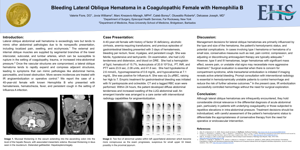
A 40-year-old female with history of factor IX deficiency, alcoholic cirrhosis, anemia requiring transfusions, and previous episodes of gastrointestinal bleeding presented with 3 days of hematemesis, bleeding per rectum, fever, myalgias, and persistent cough. She was febrile, hypotensive and tachycardic. On examination, she had LUQ tenderness and distension, and blood on DRE. She had a hemoglobin of 5g/d, hematocrit of 15.7%, leukocytosis of 20.8 10^3/uL. PT, INR, and PTT were 23.9 sec, 2.08 units, and 41.6 sec. She had hypokalemia of 2.6 mmol/L, hypomagnesemia of 0.6 mg/dL, and hypocalcemia at 7.1 mg/dL. She was positive for Influenza A. She was s/p 2u pRBC, raising her Hgb to 7. Empiric treatment for gastrointestinal bleeding was initiated with IV pantoprazole and octreotide. CT and a tagged RBC scan were performed. Within 24 hours, the patient developed diffuse abdominal tenderness and increased swelling of the LUQ abdominal wall. An emergent transfer was arranged to a care center with interventional radiology capabilities for angioembolization.
Discussion:
Management decisions for lateral oblique hematomas are primarily influenced by the type and size of the hematoma, the patient's hemodynamic status, and potential complications. In cases involving type I hematoma or hematoma of a small size, conservative measures such as rest, pain control, compression, and adjusting or discontinuing anticoagulant therapy are typically adequate.3 However, type II and III hematomas, larger hematomas with significant mass effect, severe pain, or unstable vital signs may necessitate more aggressive treatment.3 Surgical evaluation is essential when there is concern for compartment syndrome, while transarterial embolization is initiated if imaging reveals active arterial bleeding. Prompt consultation with interventional radiology is essential in hemodynamically unstable patients to control hemorrhage and reduce the risk of further adverse outcomes.4 In the present case, IR embolization successfully controlled hemorrhage without the need for surgical exploration.Conclusion:
Although lateral oblique hematomas are infrequently encountered, they hold considerable clinical relevance in the differential diagnosis of acute abdominal pain, particularly in patients with underlying coagulopathy or those subjected to repetitive elevations in intra-abdominal pressure. Treatment decisions should be individualized, with careful assessment of the patient’s hemodynamic status to differentiate the appropriateness of conservative therapy from the need for operative or endovascular intervention.
References:
-
Allen, M., & Sevensma, K. E. (2025). Rectus sheath hematoma. In StatPearls. StatPearls Publishing.https://www.ncbi.nlm.nih.gov/books/NBK519033/
-
Shimizu, T., Hanasawa, K., Yoshioka, T., Mori, T., Kajinami, T., Yokoyama, K., Sho, K., & Tani, T. (2003). Spontaneous hematoma of the lateral abdominal wall caused by a rupture of a deep circumflex iliac artery: report of two cases. Surgery today, 33(6), 475–478. https://doi.org/10.1007/s10595-002-2512-1
-
Afşin, E., & Cosgun, Z. (2021). A rare cough complication: Internal oblique muscle hematoma. Radiology Case Reports, 16(5), 1015–1018.https://doi.org/10.1016/j.radcr.2021.01.059
-
Di Pietro, S., Tiralongo, F., Desiderio, C. M., Vacirca, F., Palmucci, S., Giurazza, F., Venturini, M., & Basile, A. (2022). Efficacy of Percutaneous Transarterial Embolization in Patients with Spontaneous Abdominal Wall Hematoma and Comparison between Blind and Targeted Embolization Approaches. Journal of clinical medicine, 11(5), 1270. https://doi.org/10.3390/jcm11051270