875 posters, 25 topics, 3,440 authors, 1,061 institutions
ePostersLive by SciGen Technologies S.A. All rights reserved.
SAGES 2026 Annual Meeting Emerging Technology Session
March 25-28, 2026 | Tampa, FL, USA

P100
UNEXPECTED FINDING OF SMALL BOWEL DIVERTICULUM IN LOOP FORMATION
Poster Presenter
Authors
Affiliations
Part of Topic
Acute Care
Introduction:
Small bowel diverticulum in loop formation is an uncommon finding. The looped configuration can lead to stasis of intestinal contents, bacterial overgrowth and inflammation. We are reporting a case of a 52-year-old male with chronic non-specific gastrointestinal symptoms secondary to small bowel diverticulum in loop formation.
Case presentation:
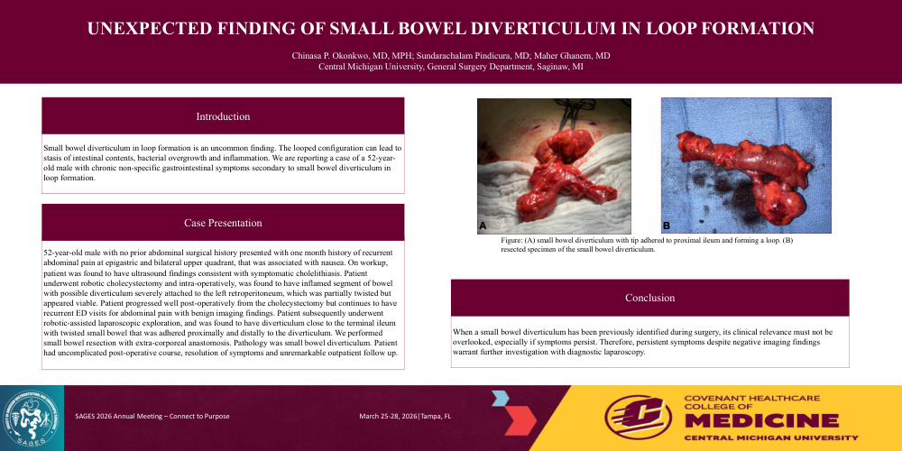
52-year-old male with no prior abdominal surgical history presented with one month history of recurrent abdominal pain at epigastric and bilateral upper quadrant, that was associated with nausea. On workup, patient was found to have ultrasound findings consistent with symptomatic cholelithiasis. Patient underwent robotic cholecystectomy and intra-operatively, was found to have inflamed segment of bowel with possible diverticulum severely attached to the left retroperitoneum, which was partially twisted but appeared viable. Patient progressed well post-operatively from the cholecystectomy but continues to have recurrent ED visits for abdominal pain with benign imaging findings. Patient subsequently underwent robotic-assisted laparoscopic exploration, and was found to have diverticulum close to the terminal ileum with twisted small bowel that was adhered proximally and distally to the diverticulum. We performed small bowel resection with extra-corporeal anastomosis. Pathology was small bowel diverticulum. Patient had uncomplicated post-operative course, resolution of symptoms and unremarkable outpatient follow up.
Discussion:
When a small bowel diverticulum has been previously identified during surgery, its clinical relevance must not be overlooked, especially if symptoms persist. Therefore, persistent symptoms despite negative imaging findings warrant further investigation with diagnostic laparoscopy.