1,267 posters, 47 videos, 13 topics, 4 sessions, 853 authors
ePostersLive by SciGen Technologies S.A. All rights reserved.
SOHO 2026 Annual Meeting
September 9 - 12, 2026 | George R. Brown Convention Center, Houston, Texas

TCL - 1326
Treatment Outcomes and Prognostic Factors in Elderly Patients with Newly Diagnosed Peripheral T-Cell Lymphoma
Poster Presenter
Authors
Part of Topic
T-Cell Lymphoma (TCL)
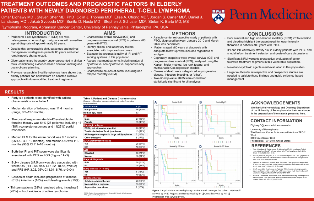
Omar Elghawy MD1 , Steven Sher MD, PhD1 Colin J. Thomas MD1 , Elise A. Chong MD1 , Jordan S. Carter MD1 , Daniel J. Landsburg MD1 , Jakub Svoboda MD1 , Sunita D. Nasta MD1 , Stephen J. Schuster MD1 , Stefan K. Barta MD, MS1 1Lymphoma Program, Abramson Cancer Center, University of Pennsylvania, Philadelphia, PA, USA TREATMENT OUTCOMES AND PROGNOSTIC FACTORS IN ELDERLY PATIENTS WITH NEWLY DIAGNOSED PERIPHERAL T-CELL LYMPHOMA • Peripheral T-cell lymphomas (PTCLs) are rare, heterogeneous hematologic malignancies with a median age at diagnosis of approximately 65 years. • Despite this demographic shift, outcomes and optimal management strategies in patients 80 years and older remain poorly characterized. • Older patients are frequently underrepresented in clinical trials, complicating evidence-based decision-making and goals-of-care planning. • Previous research in B-cell lymphomas have shown that elderly patients can benefit from an adapted curative approach with dose-modified treatment regimens. INTRODUCTION • Characterize overall survival (OS) and progression-free survival (PFS) in patients ≥80 years with PTCL • Identify clinical and laboratory factors associated with improved outcomes • Evaluate the prognostic utility of IPI and PIT scoring systems in this population • Assess treatment patterns, including rates of cytotoxic vs. non-cytotoxic vs. supportive-only approaches • Characterize causes of death, including nonrelapse mortality (NRM) AIMS • A single-center retrospective study of patients with PTCL diagnosed between January 2010 and March 2026 was performed. • Patients aged ≥80 years at diagnosis with adequate follow-up were included regardless of subtype • Coprimary endpoints were overall survival (OS) and progression-free survival (PFS), analysed using the Kaplan–Meier method, log-rank testing, and multivariable Cox regression models. • Causes of death were categorized as progressive disease, infection, bleeding, or “other”. • Two-sided p-value <0.05 were considered statistically significant for all analyses METHODS • Poor survival and high non-relapse mortality (NRM) 2 nd to infection and bleeding highlight the urgent need for lower-intensity therapies in patients ≥80 years with PTCL. • IPI and PIT effectively stratify risk in elderly patients with PTCL and should inform treatment selection and goals-of-care discussions • Significant NRM warrants prospective evaluation of bettertolerated treatment regimens in this vulnerable population. • Novel non-cytotoxic agents merit evaluation in this population. • Larger multicenter retrospective and prospective studies are needed to validate these findings and guide evidence-based management. CONCLUSIONS We thank the Hematology and Oncology Department of the University of Pennsylvania for their assistance in the preparation of the material presented here. ACKNOWLEDGEMENTS 1. Vose J, Armitage J, Weisenburger D; International T-Cell Lymphoma Project. International peripheral T-cell and natural killer/T-cell lymphoma study. J Clin Oncol. 2008;26(25):4124-4130. 2. Bellei M, Foss FM, Foyil KV, et al. The outcome of peripheral T-cell lymphoma in first complete remission with and without consolidative stem-cell transplantation. Ann Oncol. 2014;25(6):1278-1283. 3. Gallamini A, Stelitano C, Calvi R, et al. Peripheral T-cell lymphoma unspecified (PTCL-U): a new prognostic model from a retrospective multicentric clinical study. Blood. 2004;103(7):2474-2479. [PIT score] 4. Ellin F, Landström J, Jerkeman M, Relander T. Real-world data on prognostic factors and treatment in peripheral T-cell lymphomas: a study from the Swedish Lymphoma Registry. Blood. 2014;124(10):1570-1577. 5. Mead M, Cederleuf H, Björklund M, et al. Impact of comorbidity in older patients with peripheral T-cell lymphoma: an international retrospective analysis of 891 patients. Blood Adv. 2022;6(7):2120-2128. REFERENCES ElghawyO@pennmedicine.upenn.edu University of Pennsylvania The Perelman Center for Advanced Medicine TRC-2 West 3400 Civic Center Blvd Philadelphia, PA 19104, United States CONTACT INFORMATION RESULTS Table 1. Patient and Disease Characteristics Summary of baseline characteristics for 46 patients meeting inclusion criteria. ECOG, Eastern Cooperative Oncology Group; LDH, lactate dehydrogenase; PTCL, peripheral T-cell lymphoma. Characteristic n (%) Total patients 46 (100%) Median age, years 83 Sex Male 27 (59%) Female 19 (41%) Histologic subtype PTCL, not otherwise specified 20 (43%) T-follicular helper T-cell lymphoma 13 (28%) ALK-negative anaplastic large cell lymphoma 5 (11%) Other subtypes 8 (17%) ECOG performance status < 2 28 (61%) ≥ 2 18 (39%) LDH Elevated 36 (78%) Normal 10 (22%) Stage at diagnosis III/IV 28 (61%) I/II 18 (39%) Bulky disease (> 7.5 cm) Yes 6 (13%) No 40 (87%) Frontline therapy Cytotoxic chemotherapy 26 (56%) Non-cytotoxic regimen 13 (28%) Supportive care alone 7 (15%) Figure 1. Kaplan Meier curve depicting survival trends amongst the cohort. A) Overall survival by IPI B) Progression free survival by IPI C) Overall survival by PIT D) Progression free survival by PIT. • Forty-six patients were identified with patient characteristics as in Table 1. • Median duration of follow-up was 11.4 months (range, 0.2–127 months). • The overall response rate (N=42 evaluable) to frontline therapy was 64% (27 patients), including 16 (38%) complete responses and 11(26%) partial responses. • Median PFS for the entire cohort was 8.7 months (95% CI 4.8–13 months), and median OS was 11.0 months (95% CI 7.1–18 months). • Both the IPI and PIT score were significantly associated with PFS and OS (Figure 1A-D). • Bulky disease (≥7.5 cm) was also associated with worse OS (HR 3.58, 95% CI 1.22–10.52, p=0.02) and PFS (HR 3.02, 95% CI 1.04–8.76, p=0.04) • Causes of death included progression of disease (61%), infections (18%) and bleeding events (10%). • Thirteen patients (28%) remained alive, including 9 (20%) without evidence of active lymphoma.