875 posters, 25 topics, 3,440 authors, 1,061 institutions
ePostersLive by SciGen Technologies S.A. All rights reserved.
SAGES 2026 Annual Meeting Emerging Technology Session
March 25-28, 2026 | Tampa, FL, USA

P743
Standardization of Laparoscopic Peritoneal Lavage for Hinchey III Diverticulitis: A Case Report and Detailed Operative Technique
Poster Presenter
Authors
James C Rosser, Maximus L'Amour, Ascharya Balaji, Harneet Sandhu, Dylan Wong, Krishay Sridalla, Owen Burns, Luca Alessandro Arias, Zaid Elnasser, Julio Arango
Affiliations
McGill University, University at Buffalo, Jacobs School of Medicine and Biomedical Sciences, Washington and Lee University, Northwestern University Feinberg School of Medicine,
Part of Topic
New Technologies / Techniques
STANDARDIZATION OF LAPAROSCOPIC PERITONEAL LAVAGE FOR HINCHEY III DIVERTICULITIS: A CASE REPORT AND DETAILED OPERATIVE TECHNIQUE
James C Rosser, MD, FACS; Maximus L'Amour, MD, FACS; Ascharya Balaji, MD; Harneet Sandhu, MD; Dylan Wong, BS; Krishay Sridalla, BA; Owen Burns; Luca Alessandro Arias; Zaid Elnasser; Julio Arango, BS
Introduction
The use of laparoscopy peritoneal lavage (LPL) as a treatment for diverticulitis has been controversial. While initial enthusiasm has been tempered by mixed results in randomized trials, there remains a subset of patients in whom LPL may provide meaningful benefit. Its most promising role appears to be as a minimally invasive “bridge” treatment for Hinchey Class III diverticulitis. Despite its potential, there has been no detailed consensus on the precise technical execution of the procedure. This report is a case study involving a 76-year-old female patient with a Hinchey III diverticulitis and outlines a detailed operative approach, with the goal of advancing standardization and guiding future study.
Methods
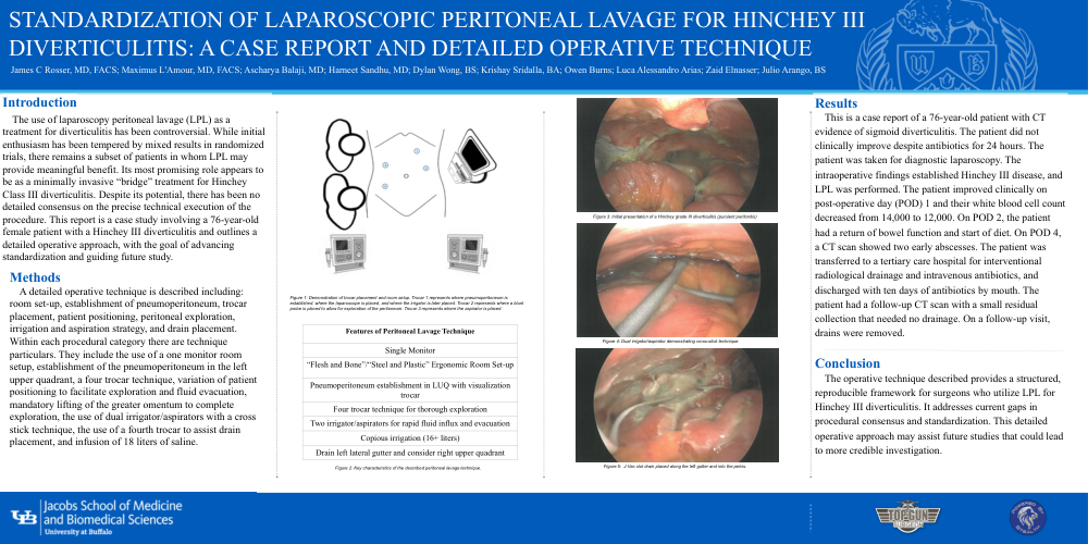
A detailed operative technique is described including: room set-up, establishment of pneumoperitoneum, trocar placement, patient positioning, peritoneal exploration, irrigation and aspiration strategy, and drain placement. Within each procedural category there are technique particulars. They include the use of a one monitor room setup, establishment of the pneumoperitoneum in the left upper quadrant, a four trocar technique, variation of patient positioning to facilitate exploration and fluid evacuation, mandatory lifting of the greater omentum to complete exploration, the use of dual irrigator/aspirators with a cross stick technique, the use of a fourth trocar to assist drain placement, and infusion of 18 liters of saline.
Figure 1: Demonstration of trocar placement and room setup. Trocar 1 represents where pneumoperitoneum is established, where the laparoscope is placed, and where the irrigator is later placed. Trocar 2 represents where a blunt probe is placed to allow for exploration of the peritoneum. Trocar 3 represents where the aspirator is placed.
|
Features of Peritoneal Lavage Technique |
|
Single Monitor |
|
“Flesh and Bone”/“Steel and Plastic” Ergonomic Room Set-up |
|
Pneumoperitoneum establishment in LUQ with visualization trocar |
|
Four trocar technique for thorough exploration |
|
Two irrigator/aspirators for rapid fluid influx and evacuation |
|
Copious irrigation (16+ liters) |
|
Drain left lateral gutter and consider right upper quadrant |
Figure 2: Key characteristics of the described peritoneal lavage technique.
Figure 3: Initial presentation of a Hinchey grade III diverticulitis (purulent peritonitis)
Figure 4: Dual irrigator/aspirator demonstrating cross-stick technique.
Figure 5: J-Vac slot drain placed along the left gutter and into the pelvis.
Results
This is a case report of a 76-year-old patient with CT evidence of sigmoid diverticulitis. The patient did not clinically improve despite antibiotics for 24 hours. The patient was taken for diagnostic laparoscopy. The intraoperative findings established Hinchey III disease, and LPL was performed. The patient improved clinically on post-operative day (POD) 1 and their white blood cell count decreased from 14,000 to 12,000. On POD 2, the patient had a return of bowel function and start of diet. On POD 4, a CT scan showed two early abscesses. The patient was transferred to a tertiary care hospital for interventional radiological drainage and intravenous antibiotics, and discharged with ten days of antibiotics by mouth. The patient had a follow-up CT scan with a small residual collection that needed no drainage. On a follow-up visit, drains were removed.
Conclusion
The operative technique described provides a structured, reproducible framework for surgeons who utilize LPL for Hinchey III diverticulitis. It addresses current gaps in procedural consensus and standardization. This detailed operative approach may assist future studies that could lead to more credible investigation.