875 posters, 25 topics, 3,440 authors, 1,061 institutions
ePostersLive by SciGen Technologies S.A. All rights reserved.
SAGES 2026 Annual Meeting Emerging Technology Session
March 25-28, 2026 | Tampa, FL, USA

P709
A RARE CASE OF PROXIMAL JEJUNAL MECKEL'S: A COURAGEOUS DISCOVERY
Poster Presenter
Authors
Asher E Lippe, Naseem Sorurbakhsh, Richard Gonzalo, Joseph Kim, Matthew Jung
Affiliations
Part of Topic
Miscellaneous / Other
A Rare Case of Proximal Jejunal Meckel’s: A Courageous Discovery
Introduction
Meckel’s diverticulum is the most common congenital anomaly of the GI tract.
Typically arises from the antimesenteric border of the distal ileum within 100 cm of the ileocecal valve.
Occurs in 2–3% of the population.
Symptomatic disease in adults is uncommon.
Proximal jejunal location is extremely rare.
This case describes proximal jejunal Meckel’s presenting as small bowel obstruction.
Case Presentation
72-year-old male with no prior abdominal surgery.
Presented with severe abdominal pain, bilious vomiting, and abdominal distension.
Laboratory evaluation revealed leukocytosis.
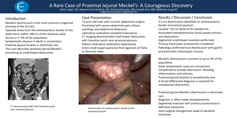
CT imaging demonstrated small bowel obstruction with transition point near proximal jejunum.
Patient underwent exploratory laparotomy.
Entire small bowel examined from ligament of Treitz to ileocecal valve.
Results / Discussion / Conclusion
3.5 cm diverticulum identified on antimesenteric border of proximal jejunum.
Located ~10 cm distal to the duodenum.
Associated mesodiverticular band caused volvulus and obstruction.
Segmental small bowel resection performed.
Primary hand-sewn anastomosis completed.
Pathology confirmed true diverticulum with gastric and pancreatic heterotopic mucosa.
Meckel’s diverticulum is present in up to 3% of the population.
Adult symptomatic cases are uncommon.
Complications include obstruction, bleeding, inflammation, and volvulus.
Proximal jejunal location is exceptionally rare.
A broad differential diagnosis is essential for unexplained obstruction.
Proximal jejunal Meckel’s diverticulum is extremely rare.
Diagnosis is often made intraoperatively.
Segmental resection with primary anastomosis is definitive treatment.
Early surgical management leads to excellent outcomes.