875 posters, 25 topics, 3,440 authors, 1,061 institutions
ePostersLive by SciGen Technologies S.A. All rights reserved.
SAGES 2026 Annual Meeting Emerging Technology Session
March 25-28, 2026 | Tampa, FL, USA

P672
Hepatogastric Fistula Secondary to Liver abscess: A Case Report
Poster Presenter
Part of Topic
Liver
Introduction
Hepatogastric fistula is a rare complication of ruptured liver abscesses, with only a small number of cases described in the literature. The diagnosis is often challenging due to vague presenting symptoms such as generalized weakness, fever, nausea, abdominal pain, and decreased appetite. We present the case of a 72-year-old female who developed a hepatogastric fistula following a liver abscess that was successfully treated with staged surgical management.
Case
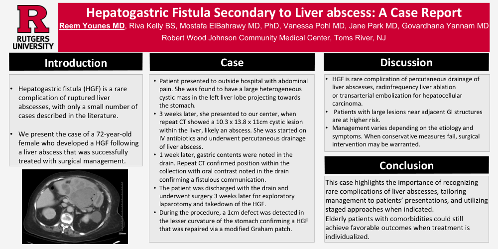
The patient initially presented to an outside hospital with abdominal pain. CT scan of the abdomen showed a large heterogeneous cystic mass in the left liver lobe projecting towards the stomach. It was biopsied and pathologic report showed fibrotic tissue with necrosis and inflammation, negative for malignancy. 3 weeks after initial presentation, she presented to our center with leg swelling. Repeat CT showed a 10.3 x 13.8 x 11cm cystic lesion within the liver, likely an abscess, and sigmoig diverticulitis with adjacent abscess 5x4.4cm. She was started on intravenous antibiotics and underwent percutaneous drainage. 7 days later, gastric contents were noted in the drain. Repeat CT confirmed appropriate position within the collection, however oral contrast was noted in the drain confirming a fistulous communication. The patient was discharged with the drain and was scheduled for surgery 3 weeks later for exploratory laparotomy, Hartmann’s procedure, and takedown of the hepatogastric fistula. A 1cm defect was detected in the lesser curvature of the stomach confirming a hepatogastric fistula that was repaired via a modified Graham patch. Six months later, she underwent colostomy takedown successfully. During the surgery, no evidence of persistent fistulous communication was found. Her postoperative course was uncomplicated, and she was discharged home tolerating a diet
Discussion
Hepatogastric fistulas have been described as a rare complication of percutaneous drainage of liver abscesses, radiofrequency liver ablation or transarterial embolization for hepatocellular carcinoma. Patients with large lesions near adjacent gastrointestinal structures are at higher risk. CT imaging and, in some cases, endoscopy are essential for diagnosis. Management varies depending on the etiology and symptoms. Some patients may be managed conservatively with antibiotics, proton pump inhibitors, and drainage. When this fails, surgical intervention may be warranted.
Conclusion
This case highlights the importance of recognizing rare complications of liver abscesses, tailoring management to patients’ presentations, and utilizing staged approaches when indicated. Elderly patients with comorbidities could still achieve favorable outcomes when treatment is individualized.