875 posters, 25 topics, 3,440 authors, 1,061 institutions
ePostersLive by SciGen Technologies S.A. All rights reserved.
SAGES 2026 Annual Meeting Emerging Technology Session
March 25-28, 2026 | Tampa, FL, USA

P667
Incidental discovery of persistent left superior vena cava during hemodialysis catheter placement: A case report
Poster Presenter
Authors
Affiliations
Part of Topic
Image Guided Interventions (Including Combined Radiologic / Endoscopic / Surgical Procedures)
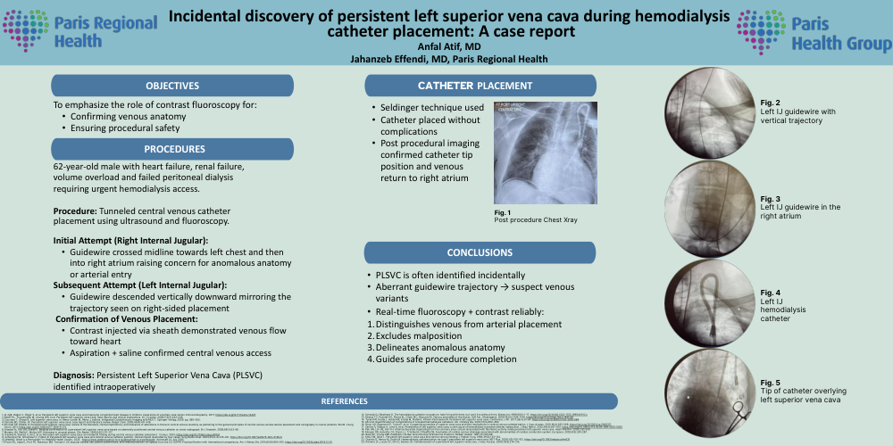
Incidental discovery of persistent left superior vena cava during hemodialysis catheter placement: A case report Anfal Atif, MD Jahanzeb Effendi, MD, Paris Regional Health 62-year-old male, ESRD, peritoneal dialysis failed due to severe LE edema. Procedure: Tunneled central venous catheter placement Initial Attempt (Right Internal Jugular): Guidewire deviated into right atrium raising concern for anomalous anatomy or arterial entry Subsequent Attempt (Left Internal Jugular): Guidewire descended vertically downward Mirrored the trajectory seen on right-sided attempt Confirmation of Venous Placement: Contrast injected via peel-pack sheath demonstrated venous flow toward heart Aspiration + saline confirmed central venous access Diagnosis: Persistent Left Superior Vena Cava (PLSVC) identified intraoperatively PROCEDURE OBJECTIVES CATHETER PLACEMENT Seldinger technique used Catheter placed without complications Post procedural imaging confirmed catheter tip position and venous return to right atrium CONCLUSIONS PLSVC is often identified incidentally Aberrant guidewire trajectory → suspect venous variants Real-time fluoroscopy + contrast reliably: 1.Distinguishes venous from arterial placement 2.Excludes malposition 3.Delineates anomalous anatomy 4.Guides safe procedure completion Fig. 1 Post procedure Chest Xray in PACU Fig. 2 Left IJ guidewire with vertical trajectory Fig. 3 Left IJ guidewire in the right atrium Fig. 4 Left IJ permcath Fig. 5 Tip of catheter overlying REFERENCES left SVC