875 posters, 25 topics, 3,440 authors, 1,061 institutions
ePostersLive by SciGen Technologies S.A. All rights reserved.
SAGES 2026 Annual Meeting Emerging Technology Session
March 25-28, 2026 | Tampa, FL, USA

P625
An Air of Uncertainty: Managing Pneumoperitoneum in Severe Pulmonary Disease Mimicking Perforated Viscus
Poster Presenter
Authors
Awinita Barpujari, Maryam Morris, Jin Y Park, Sameh Youseff, Aley E Tohamy
Affiliations
Part of Topic
Foregut
Background
Pneumoperitoneum is classically considered a surgical emergency, most often indicating perforation of a hollow viscus. However, nonsurgical pneumoperitoneum (NSP) may occur in the absence of gastrointestinal perforation, particularly in patients with underlying pulmonary pathology. Recognition of nonsurgical pneumoperitoneum is critical to avoid unnecessary operative intervention.
Case Description
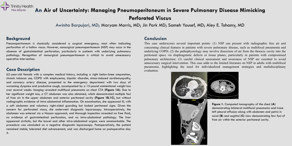
A 62-year-old female with a complex medical history, including a right below-knee amputation, chronic tobacco use, COPD with emphysema, bipolar disorder, stress-induced cardiomyopathy, and coronary artery disease, presented to the emergency department with two days of worsening dyspnea and productive cough, accompanied by a 15-pound unintentional weight loss over several weeks. Imaging revealed multifocal pneumonia on chest CTA. Due to her significant weight loss, a CT abdomen was also obtained, which demonstrated multiple foci of free air in the upper abdomen and anterior peritoneal cavity, but without radiographic evidence of intra-abdominal inflammation. On examination, she appeared ill, with a soft abdomen and voluntary right-sided guarding, but lacked peritoneal signs. Given the concern for perforated viscus, she underwent diagnostic laparoscopy. Intraoperatively, the abdomen was entered via a Hasson approach, and thorough inspection revealed no free fluid, no evidence of gastrointestinal perforation, and no intra-abdominal pathology. The liver appeared cirrhotic, but the bowel and other intra-abdominal organs were unremarkable. The procedure was concluded as a negative diagnostic laparoscopy. Postoperatively, the patient remained stable, tolerated diet advancement, and was discharged home on postoperative day 4.
Conclusion
This case underscores several important points: (1) NSP can present with radiographic free air and concerning clinical features in patients with severe pulmonary disease, such as multifocal pneumonia and underlying COPD; (2) the pathophysiology may involve dissection of air from the thoracic cavity into the peritoneal space via diaphragmatic defects or tissue planes, particularly in patients with compromised pulmonary architecture; (3) careful clinical assessment and awareness of NSP are essential to avoid unnecessary surgical intervention. This case adds to the limited literature on NSP in adults with multifocal pneumonia, highlighting the need for individualized management strategies and multidisciplinary evaluation.