875 posters, 25 topics, 3,440 authors, 1,061 institutions
ePostersLive by SciGen Technologies S.A. All rights reserved.
SAGES 2026 Annual Meeting Emerging Technology Session
March 25-28, 2026 | Tampa, FL, USA

P430
Adult Colonic Intussusception Secondary to Adenocarcinoma: A Case Report
Part of Topic
Colorectal
ADULT COLONIC INTUSSUSCEPTION SECONDARY TO ADENOCARCINOMA: CASE REPORT
Patricia Palanca, DO¹; Adelso Tejada Jackson, MD¹; Muhammad Akram²; Yaacov Bergman³; Yehuda Rosenberg³; Delcasse Joseph, MD, FASCRS¹
¹St. John’s Episcopal Hospital, ²American University of the Caribbean School of Medicine,³Touro College of Osteopathic Medicine
Introduction: Adult colonic intussusception is a rare clinical entity, accounting for approximately 1% of bowel obstructions and 5% of all intussusceptions. In contrast to pediatric intussusception, an underlying pathologic lead point is usually present in adults, with malignancy being the most frequent cause. Diagnosis is often delayed due to the nonspecific presentation, which may mimic other gastrointestinal disorders. Prompt recognition and surgical management are essential to prevent complications such as bowel ischemia and perforation. We present a case of rectosigmoid colonic intussusception secondary to adenocarcinoma, highlighting diagnostic, intraoperative, and postoperative considerations.
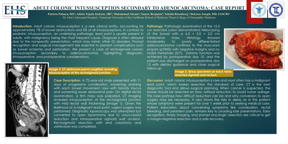
Case Description: A 72-year-old male presented with 11 days of progressive constipation, worsening straining with each bowel movement now with bloody mucus and worsening lower abdominal pain. On digital rectal examination, a firm mass was palpated. CT imaging revealed intussusception at the rectosigmoid junction with mild rectal wall thickening (Image 1). Given the likelihood of a malignant lead point, urgent surgery was performed. Diagnostic laparoscopy was attempted but converted to open laparotomy due to unsuccessful reduction and intraoperative sigmoid wall avulsion. Rectosigmoid resection with end colostomy and ureterolysis was completed.
Pathologic examination of the 15.5 cm resected colon demonstrated telescoping of the bowel with a 6.0 × 3.0 × 2.0 cm intraluminal mass. Histology showed moderately to poorly differentiated adenocarcinoma confined to the muscularis propria (pT2N0) with negative margins and no nodal metastasis (0/7). Ostomy function was achieved by postoperative day 10, and the patient was discharged on postoperative day 12 with dietary guidance and close surgical follow-up.
Discussion: Adult colonic intussusception is rare and most often has a malignant lead point, which makes resection the standard of care. CT is the best diagnostic tool and allows surgical planning. When cancer is suspected, the bowel should be resected en bloc without reduction to avoid tumor spillage. This case portrays how difficult reduction can be and why conversion to open surgery may be necessary. It also shows the risks in delay as in this patient whose symptoms were present for over 1 week prior to seeking medical care. Patient education about concerning symptoms like constipation, rectal bleeding, and persistent pain, remains key to avoiding late presentations. Early recognition, timely imaging, and prompt oncologic resection are critical to get a margin-negative resection and a safe recovery.