875 posters, 25 topics, 3,440 authors, 1,061 institutions
ePostersLive by SciGen Technologies S.A. All rights reserved.
SAGES 2026 Annual Meeting Emerging Technology Session
March 25-28, 2026 | Tampa, FL, USA

P402
Cecal Volvulus after Left Lung Lobectomy and Completion Lingulectomy: A Unique Case Report
Poster Presenter
Part of Topic
Colorectal
Cecal Volvulus after Left Lung Lobectomy and Completion Lingulectomy: A Unique Case Report
Pooja Shah, MD1, Awinita Barpujari, MD1, Franklin Burg, MD1, Tul Maya Gurung, MD1, Raj S. Lingnurkar, MD1, Maryam Morris MD1, Tiffany Schatz, MD1
1Mercy Fitzgerald Hospital, Darby, Pennsylvania 19023
Introduction
Cecal volvulus is a rare but serious condition involving the twisting of the cecum around its mesentery, often leading to closed-loop bowel obstruction. It accounts for 11–25% of all cases of colonic volvulus and is usually triggered by abnormal bowel fixation, such as adhesions, masses, or calcified lymph nodes acting as a pivot point. Gastrointestinal (GI) complications occur in roughly 1% of cardiothoracic surgeries, with ileus and pseudo-obstruction being the most frequently observed.
Case Description
A 66-year-old male with a history of diabetes, tobacco use, hyperlipidemia, and a prior left lower lobectomy for stage T1cN0M0 mucinous lung adenocarcinoma underwent the evaluation for a new, enlarging left upper lung nodule. PET-CT raised suspicion for recurrent primary lung cancer without metastasis, and biopsy confirmed adenocarcinoma. He underwent robot-assisted VATS with wedge resection, completion lingulectomy, lymph node sampling, bronchoscopy, and extensive lysis of adhesions. Intraoperatively, multiple sites of venous bleeding were noted. For concerns of coagulopathy, the patient received platelet transfusion. Postoperatively, he was extubated and admitted to the ICU.
On postoperative day 2 (POD2), the patient developed unresponsive hypotension and anemia, requiring a return to the OR for re-exploration. Active bleeding was identified in the anterior chest wall and perihilar area, along with a significant hemothorax. Hemostasis was achieved with coagulation and hemostatic agents. Postoperatively, the patient exhibited delirium and agitation, necessitating sedation with Precedex.
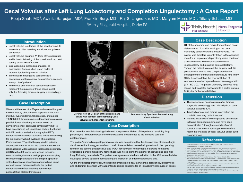
On POD3, the patient showed signs of sepsis, including tachycardia, tachypnea, leukocytosis, and abdominal distension. CT imaging revealed a markedly dilated cecum (>12 cm) with twisting of its mesentery, consistent with cecal volvulus. An emergent exploratory laparotomy confirmed the diagnosis, and an ileocecectomy with stapled side-to-side antiperistaltic anastomosis was performed. Postoperative recovery was complicated by transfusion-related acute lung injury (TRALI), requiring venovenous ECMO. The patient was eventually stabilized and discharged to a nursing facility.
Discussion
Cecal volvulus following thoracic surgery is exceptionally uncommon. Although prolonged use of muscle relaxants has been implicated in pseudo-obstruction, its link to true volvulus remains unclear. Cecal volvulus can cause ischemia, necrosis, or perforation in up to 44% of cases with similarly high mortality rates. So early diagnosis is essential. Prompt recognition and surgical intervention in postoperative patients with GI symptoms can be life-saving and should remain a critical component of postoperative care.