875 posters, 25 topics, 3,440 authors, 1,061 institutions
ePostersLive by SciGen Technologies S.A. All rights reserved.
SAGES 2026 Annual Meeting Emerging Technology Session
March 25-28, 2026 | Tampa, FL, USA

P373
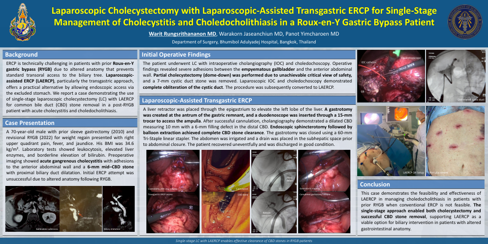
Laparoscopic Cholecystectomy with Laparoscopic-Assisted Transgastric ERCP for Single-Stage Management of Cholecystitis and Choledocholithiasis in a Roux-en-Y Gastric Bypass Patient
Poster Presenter
Authors
Warit Rungsrithananon, Warakorn Jaseanchiun, Panot Yimcharoen
Affiliations
Part of Topic
Biliary
Laparoscopic Cholecystectomy with Laparoscopic-Assisted Transgastric ERCP for Single-Stage Management of Cholecystitis and Choledocholithiasis in a Roux-en-Y Gastric Bypass Patient
Warit Rungsrithananon MD, Warakorn Jaseanchiun MD, Panot Yimcharoen MD
Department of Surgery, Bhumibol Adulyadej Hospital, Bangkok, Thailand
Background: ERCP is technically challenging in patients with prior Roux-en-Y gastric bypass (RYGB) due to altered anatomy that prevents standard transoral access to the biliary tree. Laparoscopic-assisted ERCP (LAERCP), particularly the transgastric approach, offers a practical alternative by allowing endoscopic access via the excluded stomach. We report a case demonstrating the use of single-stage laparoscopic cholecystectomy (LC) with LAERCP for common bile duct (CBD) stone removal in a post-RYGB patient with acute cholecystitis and choledocholithiasis.
Case Presentation: A 70-year-old male with prior sleeve gastrectomy (2010) and revisional RYGB (2022) for weight regain presented with right upper quadrant pain, fever, and jaundice. His BMI was 34.6 kg/m². Laboratory tests showed leukocytosis, elevated liver enzymes, and borderline elevation of bilirubin. Preoperative imaging showed acute gangrenous cholecystitis with adhesions to the anterior abdominal wall and a 6-mm mid–CBD stone with proximal biliary duct dilatation. Initial ERCP attempt was unsuccessful due to altered anatomy following RYGB. The patient underwent LC with intraoperative cholangiography (IOC) and choledochoscopy. Operative findings revealed severe adhesions between the empyematous gallbladder and the anterior abdominal wall. Partial cholecystectomy (dome-down) was performed due to unachievable critical view of safety, and a 7-mm cystic duct stone was removed. Laparoscopic IOC and choledochoscopy demonstrated complete obliteration of the cystic duct. The procedure was subsequently converted to LAERCP. A liver retractor was placed through the epigastrium to elevate the left lobe of the liver. A gastrotomy was created at the antrum of the gastric remnant, and a duodenoscope was inserted through a 15-mm trocar to access the ampulla. After successful cannulation, cholangiography demonstrated a dilated CBD measuring 10 mm with a 6-mm filling defect in the distal CBD. Endoscopic sphincterotomy followed by balloon extraction achieved complete CBD stone clearance. The gastrotomy was closed using a 60-mm Tri-Staple linear stapler. The abdomen was irrigated and a drain was placed in the subhepatic space prior to abdominal closure. The patient recovered uneventfully and was discharged in good condition.
Conclusion: This case demonstrates the feasibility and effectiveness of LAERCP in managing choledocholithiasis in patients with prior RYGB when conventional ERCP is not feasible. The single-stage approach enabled both cholecystectomy and successful CBD stone removal, supporting LAERCP as a viable option for biliary intervention in patients with altered gastrointestinal anatomy.