119 posters, 6 topics, 524 authors, 243 institutions
ePostersLive by SciGen Technologies S.A. All rights reserved.
RCR Global AI Conference 2026
29-30 June, 2026 | QEII Centre, Westminster

123
<p>Enhancing Diagnostic Accuracy in Neurology: A Deep Learning Framework for Distinguishing Multiple Sclerosis from Cerebral Small Vessel Disease on Routine Non-Contrast Brain MRI</p>
Poster Presenter
Authors
Elham Keshavarz, Kavoos Firouznia, Mehran Arab Ahmadi, Hassan Hashemi, Melika Boroomand-Saboor, Rassa Ghavami Modegh, Haned Dashti, Masoumeh Gity
Affiliations
Tehran University Of Medical Sciences, Shahid Beheshti University Of Medical Sciences, Department of Computer Engineering, Sharif University of Technology
Part of Topic
Transforming practice and leadership: pilot or test data on implementation of AI into clinical practice, clinical feedback or patient perspectives
Enhancing Diagnostic Accuracy in Neurology: A Deep Learning Framework for Distinguishing Multiple Sclerosis from Cerebral Small Vessel Disease on Routine Non-Contrast Brain MRI
Kavoos. Firouznia, Mehran. Arab Ahmadi, Hassan. Hashemi, Elham. Keshawarz, Melika. Boroomand-Saboor, Rassa. Ghavami Modegh, Hamed. Dashti, M. Gity. Presenter: Elham Keshawarz
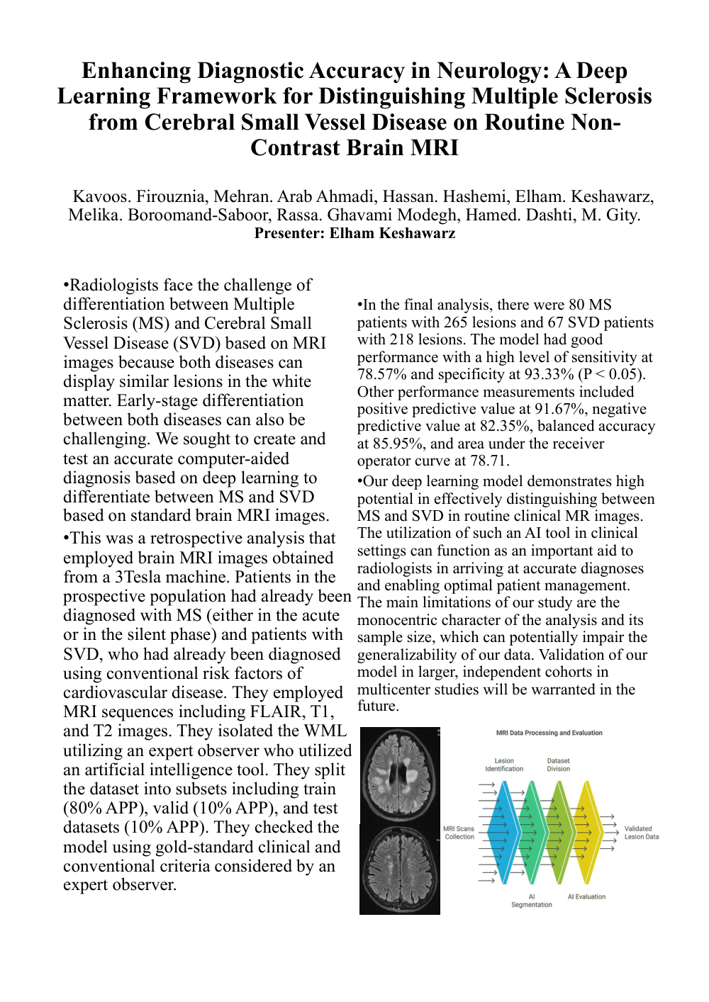
• Radiologists face the challenge of differentiation between Multiple Sclerosis (MS) and Cerebral Small Vessel Disease (SVD) based on MRI images because both diseases can display similar lesions in the white matter. Early-stage differentiation between both diseases can also be challenging. We sought to create and test an accurate computer-aided diagnosis based on deep learning to differentiate between MS and SVD based on standard brain MRI images. • This was a retrospective analysis that employed brain MRI images obtained from a 3Tesla machine. Patients in the prospective population had already been diagnosed with MS (either in the acute or in the silent phase) and patients with SVD, who had already been diagnosed using conventional risk factors of cardiovascular disease. They employed MRI sequences including FLAIR, T1, and T2 images. They isolated the WML utilizing an expert observer who utilized an artificial intelligence tool. They split the dataset into subsets including train (80% APP), valid (10% APP), and test datasets (10% APP). They checked the model using gold-standard clinical and conventional criteria considered by an expert observer. • In the final analysis, there were 80 MS patients with 265 lesions and 67 SVD patients with 218 lesions. The model had good performance with a high level of sensitivity at 78.57% and specificity at 93.33% (P < 0.05). Other performance measurements included positive predictive value at 91.67%, negative predictive value at 82.35%, balanced accuracy at 85.95%, and area under the receiver operator curve at 78.71. • Our deep learning model demonstrates high potential in effectively distinguishing between MS and SVD in routine clinical MR images. The utilization of such an AI tool in clinical settings can function as an important aid to radiologists in arriving at accurate diagnoses and enabling optimal patient management. The main limitations of our study are the monocentric character of the analysis and its sample size, which can potentially impair the generalizability of our data. Validation of our model in larger, independent cohorts in multicenter studies will be warranted in the future