296 posters, 7 videos, 13 audios, 14 topics, 10 sessions, 1,019 authors, 260 institutions
ePostersLive by SciGen Technologies S.A. All rights reserved.
RCOphth Annual Congress 2026
18 - 21 May, 2026 | Manchester Central, Manchester

P035
Bilateral Traumatic Rupture of Bruch's Membrane from Low impact Head Trauma
Poster Presenter
Authors
Part of Topic
Case reports
PURPOSE
To describe a case of profound bilateral macular destruction following an apparently low‐velocity syncopal fall in a young adult. To explore differential diagnoses when retinal injury severity is grossly disproportionate to the reported mechanism, with emphasis on traumatic choroidal/Bruch’s rupture versus self‐inflicted high‐powered laser maculopathy.
To highlight the role of multimodal imaging and systemic work‐up, for diagnostic confirmation.
INTRODUCTION
Bruch’s membrane is a thin, pentalaminar layer between the retinal pigment epithelium and choriocapillaris that supports adhesion and metabolic exchange. Traumatic choroidal rupture is a blunt‐trauma injury involving the RPE, Bruch’s membrane, and choriocapillaris, typically appearing as a white or yellow crescent‐shaped lesion concentric to the optic disc. It usually follows high‐energy orbital trauma, most often in young men after sport, assault, or projectile injury.
Bilateral traumatic choroidal rupture is exceptionally uncommon and is usually associated with severe craniofacial injury. In the Eye Injury Vitrectomy Study, a 20-year database analysis, traumatic choroidal rupture accounted for 4.8% of traumatic choroidal injuries and had an unfavourable outcome rate of 6.8% (1).
When ocular damage is far more severe than the reported mechanism, clinicians must consider either occult systemic fragility or an incomplete history. We describe a 36‐year‐old man with irreversible bilateral macular destruction after a low‐velocity syncopal fall, a presentation that prompted multidisciplinary tertiary reviews and remained a diagnostic challenge.
CASE PRESENTATION
Week 0 — Precipitating Event
COVID-19 illness → syncopal fall → frontal head impact against bath rim
Admitted in hospital unable to see; progressive "patchy" bilateral central visual loss over following days Associated mild headaches, periocular aching, and photosensitivity
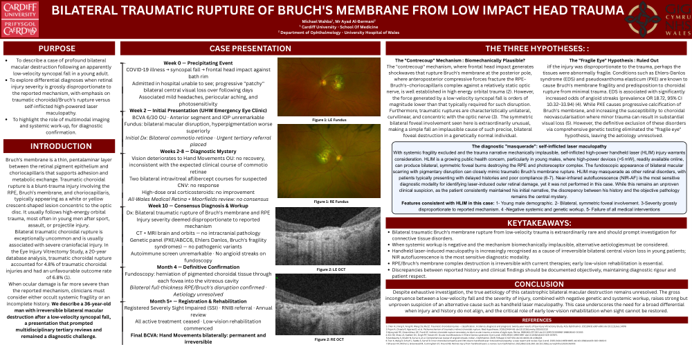
Week 2 — Initial Presentation (UHW Emergency Eye Clinic) BCVA 6/30 OU · Anterior segment and IOP unremarkable Fundus: bilateral macular disruption, hyperpigmentation worse superiorly
Initial Dx: Bilateral commotio retinae · Urgent tertiary referral placed
Weeks 2-8 — Diagnostic Mystery
Vision deteriorates to Hand Movements OU: no recovery, inconsistent with the expected clinical course of commotio retinae
Two bilateral intravitreal aflibercept courses for suspected CNV: no response
High-dose oral corticosteroids: no improvement All-Wales Medical Retina + Moorfields review: no consensus Week 10 — Consensus Diagnosis & Workup
Dx: Bilateral traumatic rupture of Bruch's membrane and RPE Injury severity deemed disproportionate to reported mechanism
CT + MRI brain and orbits — no intracranial pathology Genetic panel (PXE/ABCC6, Ehlers Danlos, Bruch's fragility syndromes) — no pathogenic variants Autoimmune screen unremarkable · No angioid streaks on fundoscopy
Month 4 — Definitive Confirmation
Fundoscopy: herniation of pigmented choroidal tissue through each fovea into the vitreous cavity
Bilateral full-thickness RPE/Bruch's disruption confirmed · Aetiology unresolved
Month 5+ — Registration & Rehabilitation Registered Severely Sight Impaired (SSI) · RNIB referral · Annual review
All active treatment ceased · Low-vision rehabilitation commenced
Final BCVA: Hand Movements bilaterally: permanent and irreversible
The "Contrecoup" Mechanism : Biomechanically Plausible?
The "contrecoup" mechanism, where frontal head impact generates shockwaves that rupture Bruch's membrane at the posterior pole, where anteroposterior compressive forces fracture the RPE– Bruch's–choriocapillaris complex against a relatively static optic nerve, is well established in high energy orbital trauma (2). However, the force generated by a low-velocity syncopal fall is orders of magnitude lower than that typically required for such disruption. Furthermore, traumatic ruptures are characteristically unilateral, curvilinear, and concentric with the optic nerve (3). The symmetric bilateral foveal involvement seen here is extraordinarily unusual, making a simple fall an implausible cause of such precise, bilateral foveal destruction in a genetically normal individual.
The "Fragile Eye" Hypothesis : Ruled Out
iIf the injury was disproportionate to the trauma, perhaps the tissues were abnormally fragile. Conditions such as Ehlers-Danlos syndrome (EDS) and pseudoxanthoma elasticum (PXE) are known to cause Bruch's membrane fragility and predisposition to choroidal rupture from minimal trauma. EDS is associated with significantly increased odds of angioid streaks (prevalence OR 18.72, 95% CI 10.32–33.94) (4). While PXE causes progressive calcification of Bruch's membrane, and increasing the susceptibility to choroidal neovascularisation where minor trauma can result in substantial visual loss (5). However, the definitive exclusion of these disorders via comprehensive genetic testing eliminated the "fragile eye" hypothesis, leaving the aetiology unresolved.
The diagnostic "masquerade": self-inflicted laser maculopathy
With systemic fragility excluded and the trauma narrative mechanically implausible, self-inflicted high-power handheld laser (HLIM) injury warrants consideration. HLIM is a growing public health concern, particularly in young males, where high-power devices (>5 mW), readily available online, can produce bilateral, symmetric foveal burns destroying the RPE and photoreceptor complex. The fundoscopic appearance of bilateral macular scarring with pigmentary disruption can closely mimic traumatic Bruch's membrane rupture. HLIM may masquerade as other retinal disorders, with patients typically presenting with delayed histories and poor compliance (6-7). Near-infrared autofluorescence (NIR-AF) is the most sensitive diagnostic modality for identifying laser-induced outer retinal damage, yet it was not performed in this case. While this remains an unproven clinical suspicion, as the patient consistently maintained his initial narrative, the discrepency between his history and the objective pathology remains the central mystery.
Features consistent with HLIM in this case: 1- Young male demographic. 2- Bilateral, symmetric foveal involvement. 3-Severity grossly disproportionate to reported mechanism. 4 -Negative systemic and genetic workup. 5- Failure of all medical interventions
KEYTAKEAWAYS:
Bilateral traumatic Bruch's membrane rupture from low-velocity trauma is extraordinarily rare and should prompt investigation for connective tissue disorders.
When systemic workup is negative and the mechanism biomechanically implausible, alternative aetiologiesmust be considered. Handheld laser-induced maculopathy is increasingly recognised as a cause of irreversible bilateral central vision loss in young patients; NIR autofluorescence is the most sensitive diagnostic modality.
RPE/Bruch's membrane complex destruction is irreversible with current therapies; early low-vision rehabilitation is essential. Discrepancies between reported history and clinical findings should be documented objectively, maintaining diagnostic rigour and patient respect.
CONCLUSION
Despite exhaustive investigation, the true aetiology of this catastrophic bilateral macular destruction remains unresolved. The gross incongruence between a low-velocity fall and the severity of injury, combined with negative genetic and systemic workup, raises strong but unproven suspicion of an alternative cause such as handheld laser maculopathy. This case underscores the need for a broad differential when injury and history do not align, and the critical role of early low-vision rehabilitation when sight cannot be restored.