296 posters, 7 videos, 13 audios, 14 topics, 10 sessions, 1,019 authors, 260 institutions
ePostersLive by SciGen Technologies S.A. All rights reserved.
RCOphth Annual Congress 2026
18 - 21 May, 2026 | Manchester Central, Manchester

P028
A Newly Recognised Vitelliform Pattern: Multifocal Vitelliform Paravascular Retinopathy
Poster Presenter
Authors
Part of Topic
Case reports
Video
Audio
Introduction
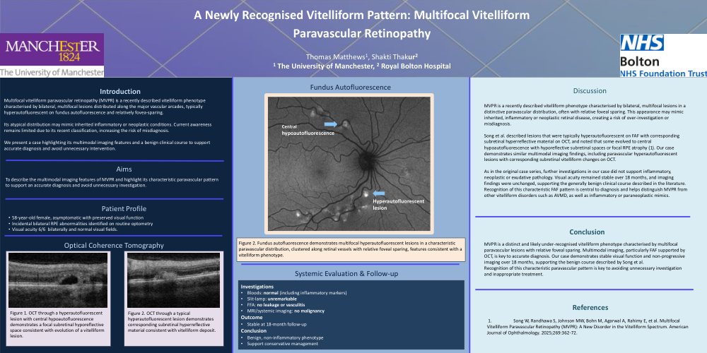
Multifocal vitelliform paravascular retinopathy (MVPR) is a recently described vitelliform phenotype characterised by bilateral, multifocal lesions distributed along the major vascular arcades, typically hyperautofluorescent on fundus autofluorescence and relatively fovea-sparing.
Its atypical distribution may mimic inherited inflammatory or neoplastic conditions. Current awareness remains limited due to its recent classification, increasing the risk of misdiagnosis.
We present a case highlighting its multimodal imaging features and a benign clinical course to support accurate diagnosis and avoid unnecessary intervention.
Aims
To describe the multimodal imaging features of MVPR and highlight its characteristic paravascular pattern to support an accurate diagnosis and avoid unnecessary investigation.
Systemic Evaluation & Follow-up
Investigations
Bloods: normal (including inflammatory markers)
Slit-lamp: unremarkable
FFA: no leakage or vasculitis
MRI/systemic imaging: no malignancy
Outcome
Stable at 18-month follow-up
Conclusion
Benign, non-inflammatory phenotype
Support conservative management
Discussion
MVPR is a recently described vitelliform phenotype characterised by bilateral, multifocal lesions in a distinctive paravascular distribution, often with relative foveal sparing. This appearance may mimic inherited, inflammatory or neoplastic retinal disease, creating a risk of over-investigation or misdiagnosis.
Song et al. described lesions that were typically hyperautofluorescent on FAF with corresponding subretinal hyperreflective material on OCT, and noted that some evolved to central hypoautofluorescence with hyporeflective subretinal spaces or focal RPE atrophy (1). Our case demonstrates similar multimodal imaging findings, including paravascular hyperautofluorescent lesions with corresponding subretinal vitelliform changes on OCT.
As in the original case series, further investigations in our case did not support inflammatory, neoplastic or exudative pathology. Visual acuity remained stable over 18 months, and imaging findings were unchanged, supporting the generally benign clinical course described in the literature. Recognition of this characteristic FAF pattern is central to diagnosis and helps distinguish MVPR from other vitelliform disorders such as AVMD, as well as inflammatory or paraneoplastic mimics.
Conclusion
MVPR is a distinct and likely under-recognised vitelliform phenotype characterised by multifocal paravascular lesions with relative foveal sparing. Multimodal imaging, particularly FAF supported by OCT, is key to accurate diagnosis. Our case demonstrates stable visual function and non-progressive imaging over 18 months, supporting the benign course described by Song et al.
Recognition of this characteristic paravascular pattern is key to avoiding unnecessary investigation and inappropriate treatment.
References
1. Song W, Randhawa S, Johnson MW, Bohn M, Agarwal A, Rahimy E, et al. Multifocal Vitelliform Paravascular Retinopathy (MVPR): A New Disorder in the Vitelliform Spectrum. American Journal of Ophthalmology. 2025;269:362-72.