296 posters, 7 videos, 13 audios, 14 topics, 10 sessions, 1,019 authors, 260 institutions
ePostersLive by SciGen Technologies S.A. All rights reserved.
RCOphth Annual Congress 2026
18 - 21 May, 2026 | Manchester Central, Manchester

P034
Beyond the Usual Demographic: Planum Sphenoidale Meningioma in a Young Man
Poster Presenter
Part of Topic
Case reports
Audio
Beyond the Usual Demographic: Planum Sphenoidale Meningioma in a Young Man
Lazar Krstic MMedSci (Dist); Ian Carmody BMBS (Hons); Melissa E Hickinbottom BMedSci; Laurence J Glancz FRCS (SN); Arpita Chattopadhyay FRCR; Anna M Gruener FRCOphth
Background
Meningiomas are the most common non-glial primary central nervous system tumours, and are most often diagnosed in middle-aged women. In younger adults, occurrence should prompt evaluation for an underlying genetic predisposition - particularly neurofibromatosis type 2. Sporadic cases without a genetic predisposition do occur and may present atypically.
Planum sphenoidale meningiomas represent a small subset of anterior skull base meningiomas and typically cause slowly progressive visual decline due to optic nerve compression. In younger patients, such lesions may follow a more aggressive course and display non-classical radiological features.
Case
A 22-year-old white man presented to the ophthalmic emergency department with a two-month history of painless, progressive blurred vision, predominantly in the left eye, first noticed acutely during sports training. The deficit later became bilateral, with the left remaining more severely affected. There was a family history of sarcoidosis.
BCVA — RE 6/24, LE HM
CV — RE 8/17 slow, LE 0/17
Anterior segment — unremarkable
Pupils — L RAPD
Posterior segment — BE mild optic disc pallor + superimposed mild oedema
IOP — normal
OCT — BE thinning of the peripapillary RNFL and macular GCL, more marked in the left eye.
HVF— RE central/paracentral defects, LE marked field loss.
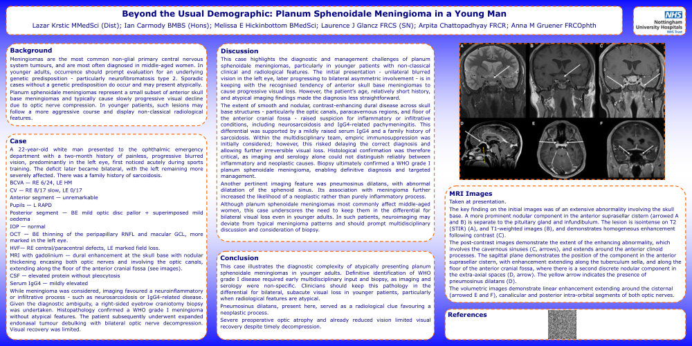
MRI with gadolinium — dural enhancement at the skull base with nodular thickening encasing both optic nerves and involving the optic canals, extending along the floor of the anterior cranial fossa (see images).
CSF — elevated protein without pleocytosis
Serum IgG4 — mildly elevated
While meningioma was considered, imaging favoured a neuroinflammatory or infiltrative process - such as neurosarcoidosis or IgG4-related disease. Given the diagnostic ambiguity, a right-sided eyebrow craniotomy biopsy was undertaken. Histopathology confirmed a WHO grade I meningioma without atypical features. The patient subsequently underwent expanded endonasal tumour debulking with bilateral optic nerve decompression. Visual recovery was limited.
Discussion
This case highlights the diagnostic and management challenges of planum sphenoidale meningiomas, particularly in younger patients with non-classical clinical and radiological features. The initial presentation - unilateral blurred vision in the left eye, later progressing to bilateral asymmetric involvement - is in keeping with the recognised tendency of anterior skull base meningiomas to cause progressive visual loss. However, the patient’s age, relatively short history, and atypical imaging findings made the diagnosis less straightforward.
The extent of smooth and nodular, contrast-enhancing dural disease across skull base structures - particularly the optic canals, paracavernous regions, and floor of the anterior cranial fossa - raised suspicion for inflammatory or infiltrative conditions, including neurosarcoidosis and IgG4-related pachymeningitis. This differential was supported by a mildly raised serum IgG4 and a family history of sarcoidosis. Within the multidisciplinary team, empiric immunosuppression was initially considered; however, this risked delaying the correct diagnosis and allowing further irreversible visual loss. Histological confirmation was therefore critical, as imaging and serology alone could not distinguish reliably between inflammatory and neoplastic causes. Biopsy ultimately confirmed a WHO grade I planum sphenoidale meningioma, enabling definitive diagnosis and targeted management.
Another pertinent imaging feature was pneumosinus dilatans, with abnormal dilatation of the sphenoid sinus. Its association with meningioma further increased the likelihood of a neoplastic rather than purely inflammatory process.
Although planum sphenoidale meningiomas most commonly affect middle-aged women, this case underscores the need to keep them in the differential for bilateral visual loss even in younger adults. In such patients, neuroimaging may deviate from typical meningioma patterns and should prompt multidisciplinary discussion and consideration of biopsy.
Conclusion
This case illustrates the diagnostic complexity of atypically presenting planum sphenoidale meningiomas in younger adults. Definitive identification of WHO grade I disease required early multidisciplinary input and biopsy, as imaging and serology were non-specific. Clinicians should keep this pathology in the differential for bilateral, subacute visual loss in younger patients, particularly when radiological features are atypical.
Pneumosinus dilatans, present here, served as a radiological clue favouring a neoplastic process.
Severe preoperative optic atrophy and already reduced vision limited visual recovery despite timely decompression.
MRI Images
Taken at presentation.
The key finding on the initial images was of an extensive abnormality involving the skull base. A more prominent nodular component in the anterior suprasellar cistern (arrowed A and B) is separate to the pituitary gland and infundibulum. The lesion is isointense on T2 (STIR) (A), and T1-weighted images (B), and demonstrates homogeneous enhancement following contrast (C).
The post-contrast images demonstrate the extent of the enhancing abnormality, which involves the cavernous sinuses (C, arrows), and extends around the anterior clinoid processes. The sagittal plane demonstrates the position of the component in the anterior suprasellar cistern, with enhancement extending along the tuberculum sella, and along the floor of the anterior cranial fossa, where there is a second discrete nodular component in the extra-axial spaces (D, arrow). The yellow arrow indicates the presence of pneumosinus dilatans (D).
The volumetric images demonstrate linear enhancement extending around the cisternal (arrowed E and F), canalicular and posterior intra-orbital segments of both optic nerves.