301 posters, 50 videos, 13 topics, 13 sessions, 734 authors, 193 institutions
ePostersLive by SciGen Technologies S.A. All rights reserved.
ISMICS 2026 Annual Meeting
10 - 13 June, 2026 | Miami, Florida

P96
Robotic, Hybrid And Endovascular Approaches To Facilitate Management Of Vascular Rings
Poster Presenter
Part of Topic
Pediatric - Cardiac, Cardiovascular & Thoracic
Robotic, Hybrid and Endovascular Approaches to Facilitate Management of Vascular Rings
Danielle R. Pitchon BS1-2, John A. Kucera MD1,3, Seth E.M. Wolf MD1,3, Rebekah R. Boyd MD1,3, Lindsey M. Reynolds BS1-2, Akosua D. Odei BA1-2, Kaitlin C. Bevers MD3, Joseph W. Turek MD PhD MBA1,3, Douglas M. Overbey MD MPH1,3
1Duke Congenital Heart Surgery Research and Training Lab, Durham, NC, USA; 2Duke University School of Medicine, Durham, NC, USA; 3Department of Surgery, Duke University Medical Center, Durham, NC, USA
BACKGROUND:
Vascular rings result from aberrant development of the great vessels and their branches. Although typically asymptomatic, they may result in dyspnea, stridor, and dysphagia lusoria. Although historically treated via thoracotomy, appropriately selected patients may benefit from minimally invasive approaches. We describe the successful management of four patients with symptomatic vascular rings by robot-assisted division or resection of aberrant vessels.
METHODS:
Four patients with symptomatic vascular rings were evaluated to determine candidacy for minimally invasive intervention. All patients presented with dysphagia and dyspnea, one reported chest pain, two had suspected asthma histories, and one exhibited stridor. Mean (range) age was 22 years (13–29), height was 158.5 cm (151.2–170.2), weight was 68.3 kg (47.8–90.9), and body mass index was 26.73 kg/m2 (20.91–31.38).
Cross sectional imaging, bronchoscopy, and esophagogastroduodenoscopy confirmed compression of aerodigestive structures. All patients met criteria to accommodate the robot (at least 6cm from mediastinum to lateral chest wall).
Two patients had right-sided aortic arches, left ligamentum arteriosum with anomalous retroesophageal left subclavian arteries, and Kommerell diverticulums. One patient had a double aortic arch, and one patient had a right-sided aortic arch with Kommerell diverticulum. The first two patients underwent robot-assisted division of the aberrant subclavian artery and ligamentum arteriosum, the third patient underwent a staged hybrid approach with robot-assisted double arch division followed by endovascular repair (TEVAR), and the fourth patient underwent TEVAR with subclavian embolization followed by robot-assisted resection of aberrant subclavian artery.
RESULTS:
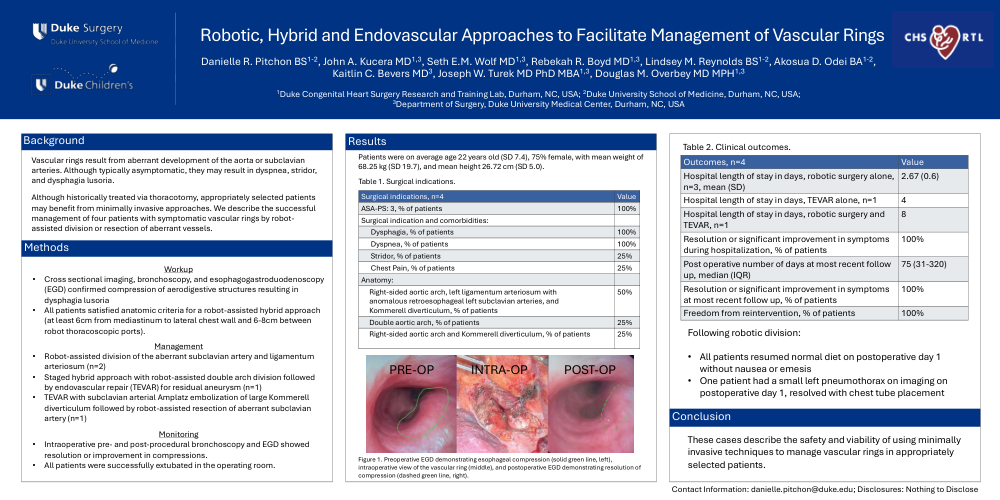
In all cases, intra-operative esophagogastroduodenoscopy confirmed resolution of anatomic compression (Figure 1). Average (SD) length of hospital stay for patients undergoing robot-assisted surgery alone was 2.67 days (0.6). All patients resumed a normal diet on post operative day 1 without nausea or emesis. All patients reported mitigation or significant improvement in symptoms during hospitalization and at most recent follow up (median 75 days; IQR: 31-320).
CONCLUSIONS:
These cases describe the safety and viability of using minimally invasive techniques to manage vascular rings in appropriately selected patients.