301 posters, 50 videos, 13 topics, 13 sessions, 734 authors, 193 institutions
ePostersLive by SciGen Technologies S.A. All rights reserved.
ISMICS 2026 Annual Meeting
10 - 13 June, 2026 | Miami, Florida

D32
Robotic-assisted Left Anterior Descending Coronary Artery Unroofing Of A Myocardial Bridge
Poster Presenter
Part of Topic
Coronary Revascularization
Video
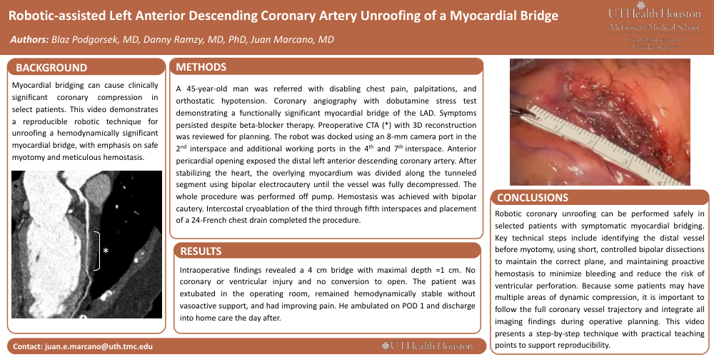
BACKGROUND: Myocardial bridging can cause clinically significant coronary compression in select patients. This video demonstrates a reproducible robotic technique for unroofing a hemodynamically significant myocardial bridge, with emphasis on safe myotomy and meticulous hemostasis.
METHODS: A 45-year-old man was referred with disabling chest pain, palpitations, and orthostatic hypotension. Coronary angiography with dobutamine stress test demonstrating a functionally significant myocardial bridge of the LAD. Symptoms persisted despite beta-blocker therapy. Preoperative CTA (*) with 3D reconstruction was reviewed for planning. The robot was docked using an 8-mm camera port in the 2nd interspace and additional working ports in the 4th and 7th interspace. Anterior pericardial opening exposed the distal left anterior descending coronary artery. After stabilizing the heart, the overlying myocardium was divided along the tunneled segment using bipolar electrocautery until the vessel was fully decompressed. The whole procedure was performed off pump. Hemostasis was achieved with bipolar cautery. Intercostal cryoablation of the third through fifth interspaces and placement of a 24-French chest drain completed the procedure.
RESULTS: Intraoperative findings revealed a 4 cm bridge with maximal depth ≈1 cm. No coronary or ventricular injury and no conversion to open. The patient was extubated in the operating room, remained hemodynamically stable without vasoactive support, and had improving pain. He ambulated on POD 1 and discharge into home care the day after.
CONCLUSIONS: Robotic coronary unroofing can be performed safely in selected patients with symptomatic myocardial bridging. Key technical steps include identifying the distal vessel before myotomy, using short, controlled bipolar dissections to maintain the correct plane, and maintaining proactive hemostasis to minimize bleeding and reduce the risk of ventricular perforation. Because some patients may have multiple areas of dynamic compression, it is important to follow the full coronary vessel trajectory and integrate all imaging findings during operative planning. This video presents a step-by-step technique with practical teaching points to support reproducibility.