206 posters, 13 topics, 5 sessions, 713 authors, 294 institutions
ePostersLive by SciGen Technologies S.A. All rights reserved.
AAPS 104th Annual Meeting
May 2-5, 2026 | Lihue, HI

D43
Influence Of Vomer Flaps Or Lateral Releasing Incisions During Primary Palatoplasty On Midface Growth: A Meta-analysis
Poster Presenter
Part of Topic
Craniofacial/Peds
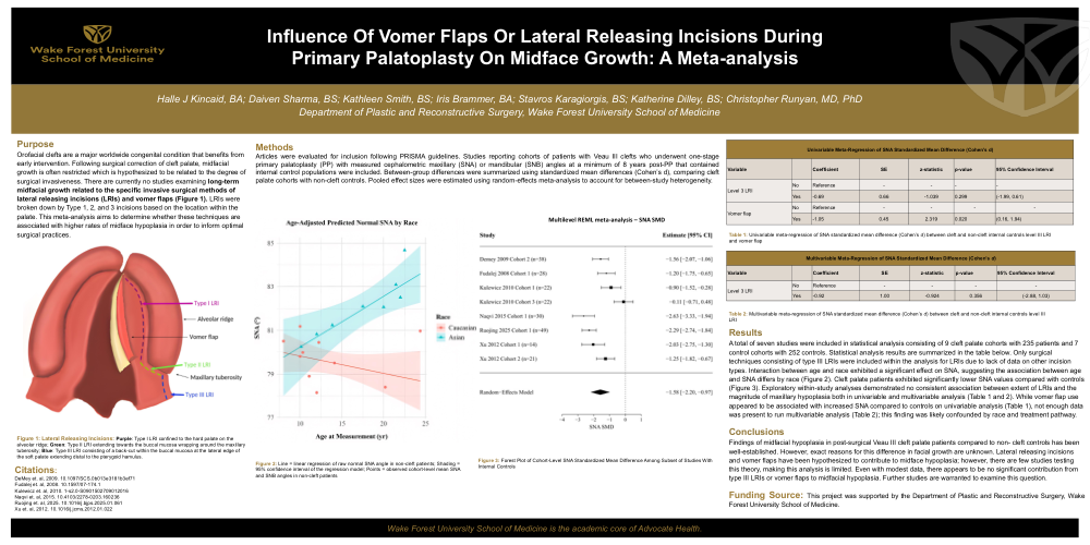
Purpose: Orofacial clefts are a major worldwide congenital condition that benefits from early intervention. Following surgical correction of cleft palate, midfacial growth is often restricted which is hypothesized to be related to the degree of surgical invasiveness. There are currently no studies examining long-term midfacial growth related to the specific invasive surgical methods of lateral releasing incisions (LRIs) (Figure 1) and vomer flaps. LRIs were broken down by Type 1, 2, and 3 incisions based on the location within the palate. This meta-analysis aims to determine whether these techniques are associated with higher rates of midface hypoplasia in order to inform optimal surgical practices.
Methods: Articles were evaluated for inclusion following PRISMA guidelines. Studies reporting cohorts of patients with Veau III clefts who underwent one-stage primary palatoplasty (PP) with measured cephalometric maxillary (SNA) or mandibular (SNB) angles at a minimum of 8 years post-PP that contained internal control populations were included. Between-group differences were summarized using standardized mean differences (Cohen’s d), comparing cleft palate cohorts with non-cleft controls. Pooled effect sizes were estimated using random-effects meta-analysis to account for between-study heterogeneity.
Results: A total of seven studies were included in statistical analysis consisting of 9 cleft palate cohorts with 235 patients and 7 control cohorts with 252 controls. Statistical analysis results are summarized in the table below. Only surgical techniques consisting of type III LRIs were included within the analysis for LRIs due to lack of data on other incision types. Interaction between age and race exhibited a significant effect on SNA, suggesting the association between age and SNA differs by race (Figure 2). Cleft palate patients exhibited significantly lower SNA values compared with controls (Figure 3). Exploratory within-study analyses demonstrated no consistent association between extent of LRIs and the magnitude of maxillary hypoplasia both in univariable and multivariable analysis (Table 1 and 2). While vomer flap use appeared to be associated with increased SNA compared to controls on univariable analysis (Table 1), not enough data was present to run multivariable analysis (Table 2); this finding was likely confounded by race and treatment pathway.
Conclusions: Findings of midfacial hypoplasia in post-surgical Veau III cleft palate patients compared to non- cleft controls has been well-established. However, exact reasons for this difference in facial growth are unknown. Lateral releasing incisions and vomer flaps have been hypothesized to contribute to midface hypoplasia; however, there are few studies testing this theory, making this analysis is limited. Even with modest data, there appears to be no significant contribution from type III LRIs or vomer flaps to midfacial hypoplasia. Further studies are warranted to examine this question.