875 posters, 25 topics, 3,440 authors, 1,061 institutions
ePostersLive by SciGen Technologies S.A. All rights reserved.
SAGES 2026 Annual Meeting Emerging Technology Session
March 25-28, 2026 | Tampa, FL, USA

P713
Paroxysmal Hypertension and Abdominal Pain: A Case of Left Adrenal Pheochromocytoma Treated by Laparoscopy
Poster Presenter
Part of Topic
Miscellaneous / Other
INTRODUCTION
Pheochromocytomas are neuroendocrine tumors that produce catecholamines and can be of adrenal or extra-adrenal origin. The classic symptoms of pheochromocytoma are headache, palpitations, anxiety, and diaphoresis, and the tumor can appear at any age with an equal distribution between sexes. In patients with an established mutation or a hereditary syndrome, the condition may manifest at an earlier age than in those with sporadic disease. Pheochromocytoma can be associated with certain genetic syndromes such as multiple endocrine neoplasia type 2 (MEN 2), neurofibromatosis (NF), and von Hippel-Lindau (VHL) syndrome. Pheochromocytoma is diagnosed by biochemical confirmation of hormonal excess followed by anatomical localization (CT or MRI). The basis of definitive treatment is surgical resection.
CASE PRESENTATION
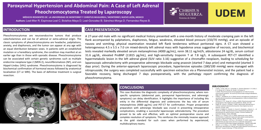
A 27-year-old male with no significant medical history presented with a one-month history of moderate cramping pain in the left flank accompanied by palpitations, diaphoresis, fatigue, weakness, elevated blood pressure (150/70 mmHg), and an episode of nausea and vomiting; physical examination revealed left flank tenderness without peritoneal signs. A CT scan showed a heterogeneous 4.5 x 5.5 x 7.0 cm mixed-density left adrenal mass with hypodense areas suggestive of necrosis, and biochemical tests revealed markedly elevated serum metanephrines (4000 pg/mL), renin 38.11 ng/ml/h, aldosterone 24 ng/dL, serum cortisol 31.6 µg/dL, elevated ProBNP (11815 pg/mL), and high-sensitivity troponin T at 7.8 ng/L. A subsequent PET-CT identified a hypermetabolic lesion in the left adrenal gland (SUV ratio 3.16) suggestive of a chromaffin neoplasm, leading to scheduling for laparoscopic adrenalectomy with preoperative adrenergic blockade using prazosin (started 7 days prior) and metoprolol (started 3 days prior). During the anterior-approach laparoscopic procedure, hypertensive episodes (180/100 mmHg) were managed with nitroprusside; the surgery was completed successfully with specimen extraction via a Pfannenstiel incision, and the patient had a favorable recovery, being discharged 7 days postoperatively, with the pathology report confirming the diagnosis of pheochromocytoma.
CONCLUSIONS
This case illustrates the diagnostic complexity of pheochromocytoma, where non-specific symptoms (abdominal pain, paroxysmal hypertension, and adrenergic symptoms) can delay identification. It highlights the importance of including this entity in the differential diagnosis and underscores the key role of serum metanephrines (4000 pg/mL) and PET-CT for confirmation. Proper preoperative preparation with adrenergic blockade was crucial in preventing intraoperative complications, enabling a successful laparoscopic adrenalectomy. The procedure proved to be safe and effective, with an optimal postoperative course and complete resolution of symptoms. This reinforces the minimally invasive approach as the gold standard for such cases when performed by experienced, multidisciplinary teams.