875 posters, 25 topics, 3,440 authors, 1,061 institutions
ePostersLive by SciGen Technologies S.A. All rights reserved.
SAGES 2026 Annual Meeting Emerging Technology Session
March 25-28, 2026 | Tampa, FL, USA

P696
Adenoma-Hemangioma Adrenal Collision Tumor Associated with Mild Autonomous Cortisol Secretion: A Case Report
Poster Presenter
Authors
Affiliations
Part of Topic
Miscellaneous / Other
Adenoma-Hemangioma Adrenal Collision Tumor Associated with Mild Autonomous Cortisol Secretion: A Case Report
Miguel A. Ortiz Rivera, MD; Frances T. Lee, MD, FACS
Department of Surgery, Southern Illinois University School of Medicine, Springfield, Illinois
Introduction
Adrenal collision tumors (ACTs) are rare masses of the adrenal
gland where two histologically distinct tumors coexist within
an adrenal gland. Due to their complex appearance on
imaging, accurate preoperative diagnosis can be challenging,
often requiring a multimodal imaging approach.
Adenoma-hemangioma ACTs have been reported very few
times in the literature. To the best of our knowledge, this is
one of two hormonally active adenoma-hemangioma ACT
reported in the literature.
Presentation
A 65-year-old female was evaluated in the endocrine surgery
clinic due to an incidentally found adrenal tumor during chest
computed tomography (CT) for cardiac scoring.
Follow-up CT of the abdomen and pelvis:
- Pre-contrast: 4 x 5.5 cm left adrenal tumor without local invasion
or calcification but with heterogeneous density.
- Arterial phase (35s): punctate foci of enhancement.
- Portal venous (70s): increasing enhancement compared to the
arterial phase.
- Delayed (4 min): Persistent nodular enhancement.
Impression: heterogenous mass concerning for
pheochromocytoma vs adrenocortical carcinoma.
Local symptoms: no
Symptoms of excess catecholamines: no
Symptoms of hypercortisolism: osteopenia
Symptoms of hyperaldosteronism: no
Physical exam: unremarkable.
Histories: Thalassemia, osteoarthritis, glaucoma, family
history of colorectal cancer.
Workup
Biochemical Profile:
Test Result Reference Range
24 hr urine free cortisol 62.6 mcg/dL <45
Dexamethasone suppression test (1g) 4.4 mcg/dL (10:04 AM) <1.8
Adrenocorticotropic Hormone 5.7pg/mL 7-63
24 hr urine catecholamines
- Epinephrine
- Norepinephrine
- Metanephrine
- Normetanephrine
Potassium 3.7mmol/L 3.5-5.1
Serum Aldosterone 5.9 ng/dL 3-35
Plasma Renin Activity 0.4 ng/mL/Hr 0.2-1.6 (supine)
DHEA-S 7 mcg/dL 24-244
Surgery & Recovery
Laparoscopic Left Adrenalectomy
- R lateral decubitus positioning, OG tube, Foley catheter.
- Veress insufflation at Palmer’s point. Ports: three 5mm ports
(paramedian supraumbilical, epigastric and left flank), and one 12mm
port (left subcostal midclavicular line),
- Splenic flexure mobilization.
- Lateral to medial mobilization of the pancreas, spleen, greater
curvature of the stomach.
- Mass identified: no desmoplastic reaction or local tissue invasion.
- The left inferior phrenic vein was traced down to its left adrenal vein
junction and renal vein termination.
- The adrenal vein was ligated twice on the stay side and a bipolar
energy device used to transect the vein on the specimen side.
- Dissection continued in the cephalad and lateral directions until the
specimen was freed & removed from the enlarged 12mm port site.
Post Operative Course
- Hemodynamics were stable during overnight observation.
- AM cortisol was low (12.1 mcg/dL, N >15).
- Follow up cosyntropin stimulation test indicated adrenal
insufficiency. (12.1 mcg/dL @ 30 min, 14.7 @ 60 min; N >18).
- A corticosteroid regimen with 10mg of hydrocortisone qAM and 5 mg
of hydrocortisone qPM was started.
- Discharge on POD 1.
- No complaints on 2 week post operative evaluation.
- The patient continues on a steroid regimen (approximately 11
months post op).
Pathology
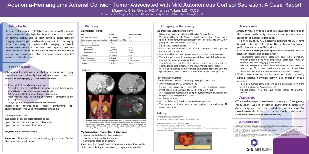
Figure 1: Preoperative imaging of the left adrenal mass. CT axial views in the portal venous
(a) and delayed (b) phases showing heterogeneous enhancement and persistent nodular
enhancement, respectively. FDG PET coronal view (c) showing mildly hypermetabolic left
adrenal activity (SUVmax 2.6, white arrow). MRI without contrast axial view (d) showing
medial high T2 signal (red arrow) with and lateral intralesional fat (yellow arrow).
Multidisciplinary Tumor Board Discussion:
- Mass more likely benign than malignant.
- Low concern for metastatic disease.
- Surveillance could be an option.
Given size, hormonally active tumor, and patient desire for
definitive pathological evaluation, surgery was elected.
1. 2. 3. 4. 5. Discussion
- Although rare, a wide gamut of ACTs have been described in
the literature with benign, metastatic, and primary adrenal
malignant components described.
- To our knowledge, <10 adenoma-hemangioma ACTs have
been described in the literature. Their potential biochemical
profile has not been well described.
- Due to their heterogeneous appearance, diagnosis of ACTs
based on imaging can be challenging
- Hemangioma components classically have early peripheral
nodular enhancement with progressive centripetal filling on
contrast enhanced imaging (CT and MRI).
- Adenoma components will be hypodense lesions with <10 HU on
non-contrast CT, or have rapid washout on the 15 min delay
phase. MRI will show a signal drop on out-of-phase T2 images.
- While surveillance can be considered for benign appearing
adrenal lesions, hormonal activity and evolution should
assessed.
- Functional tumors carry long-term risk of morbidity, even if the
patient is otherwise “asymptomatic”.
- Adrenal masses >4-6 cm have higher chance of malignant
evolution.
Conclusion
ACTs should undergo thorough workup for signs of malignancy
and function. Even in otherwise asymptomatic patients in
which malignancy has been ruled-out, consideration for
adrenalectomy should be given to hormonally active tumors
due to long-term risk of medical morbidity.