875 posters, 25 topics, 3,440 authors, 1,061 institutions
ePostersLive by SciGen Technologies S.A. All rights reserved.
SAGES 2026 Annual Meeting Emerging Technology Session
March 25-28, 2026 | Tampa, FL, USA

P652
ENCYSTED NECROSIS AFTER COSMETIC FAT GRAFTING PROCEDURE ON THE ABDOMINAL WALL MIMICKING AN ABDOMINAL WALL TUMOR: A CASE REPORT
Part of Topic
Global Surgery
INTRODUCTION:
Abdominal wall tumors are a heterogeneous group of benign and malignant neoplasms. These tumors result in the abnormal growth of abdominal muscle cells or other tissues present in the abdominal wall. The main neoplasms found are desmoid tumors and sarcomas, with fibrosarcoma being the most common subtype.
The diagnosis is made through imaging studies, and surgical resection is the primary treatment. The differential diagnosis of abdominal wall tumors is uncommon, with abdominal wall endometriosis being the main differential diagnosis.
An increasing number of cosmetic surgical procedures on the abdominal wall can lead to complications that can mimic abdominal wall tumors. Liposuction is gaining new perspectives with the development of high-definition liposuction (HD or LAD) and autologous fat grafting, which improve the definition of the abdominal wall, particularly the rectus abdominis muscle.
However, complications associated with these techniques, especially fat grafting (reinjection of fat into points on the abdominal wall), can cause inflammatory reactions and fat necrosis at the graft sites.
This condition can lead to symptoms that mimic abdominal wall tumors.
CASE DISCUSSION:
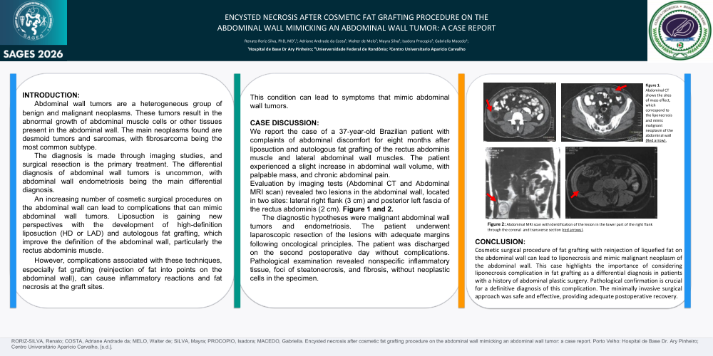
We report the case of a 37-year-old Brazilian patient with complaints of abdominal discomfort for eight months after liposuction and autologous fat grafting of the rectus abdominis muscle and lateral abdominal wall muscles. The patient experienced a slight increase in abdominal wall volume, with palpable mass, and chronic abdominal pain.
Evaluation by imaging tests (Abdominal CT and Abdominal MRI scan) revealed two lesions in the abdominal wall, located in two sites: lateral right flank (3 cm) and posterior left fascia of the rectus abdominis (2 cm). Figure 1 and 2.
The diagnostic hypotheses were malignant abdominal wall tumors and endometriosis. The patient underwent laparoscopic resection of the lesions with adequate margins following oncological principles. The patient was discharged on the second postoperative day without complications. Pathological examination revealed nonspecific inflammatory tissue, foci of steatonecrosis, and fibrosis, without neoplastic cells in the specimen.
CONCLUSION:
Cosmetic surgical procedure of fat grafting with reinjection of liquefied fat on the abdominal wall can lead to liponecrosis and mimic malignant neoplasm of the abdominal wall. This case highlights the importance of considering liponecrosis complication in fat grafting as a differential diagnosis in patients with a history of abdominal plastic surgery. Pathological confirmation is crucial for a definitive diagnosis of this complication. The minimally invasive surgical approach was safe and effective, providing adequate postoperative recovery.