875 posters, 25 topics, 3,440 authors, 1,061 institutions
ePostersLive by SciGen Technologies S.A. All rights reserved.
SAGES 2026 Annual Meeting Emerging Technology Session
March 25-28, 2026 | Tampa, FL, USA

P539
COMPARATIVE DIAGNOSTIC ACCURACY ACROSS MULTIPLE IMAGE-ENHANCED ENDOSCOPY MODALITIES FOR GASTRIC INTESTINAL METAPLASIA: A BAYESIAN BIVARIATE RANDOM-EFFECTS NETWORK META-ANALYSIS
Poster Presenter
Authors
Muhammad Mujtaba Rasool, Haneen Kamran, Asra tus Saleha Siddiqui, Muhammad Murtaza, Shameer Tahir, Wajeeha Fatima Tareen, Farah Khan
Affiliations
The Agha Khan University, Allama Iqbal Medical College, Lahore, The Aga Khan University Stadium Road Karachi, Services Institute of Medical Sciences, Lahore
Part of Topic
Flexible Endoscopy
COMPARATIVE DIAGNOSTIC ACCURACY ACROSS MULTIPLE IMAGE-ENHANCED ENDOSCOPY MODALITIES FOR GASTRIC INTESTINAL METAPLASIA: A BAYESIAN BIVARIATE RANDOM-EFFECTS NETWORK META-ANALYSIS
Introduction:
Gastric intestinal metaplasia (GIM), a precursor to gastric cancer, is often missed by standard white-light endoscopy (WLE), highlighting a critical need for advanced endoscopic imaging to improve detection and outcomes.
Methods.
We conducted a Bayesian bivariate random-effects network meta-analysis of diagnostic accuracy for gastric intestinal metaplasia (GIM). Eligible studies were prospective comparative diagnostic accuracy trials in adults, assessing endoscopic modalities against standard WLE. Interventions included image-enhanced endoscopy techniques (NBI, magnifying NBI, chromoendoscopy, optical enhancement (OE), AI-assisted modalities) and microendoscopy (CLE, pCLE). PubMed, Embase, and Scopus were searched (>2,300 records screened). Arm-level counts were extracted. Sensitivity and specificity were modeled jointly on the logit scale with correlated study-level random effects. Posterior means and 95% credible intervals (CrIs) were summarized.
Results.
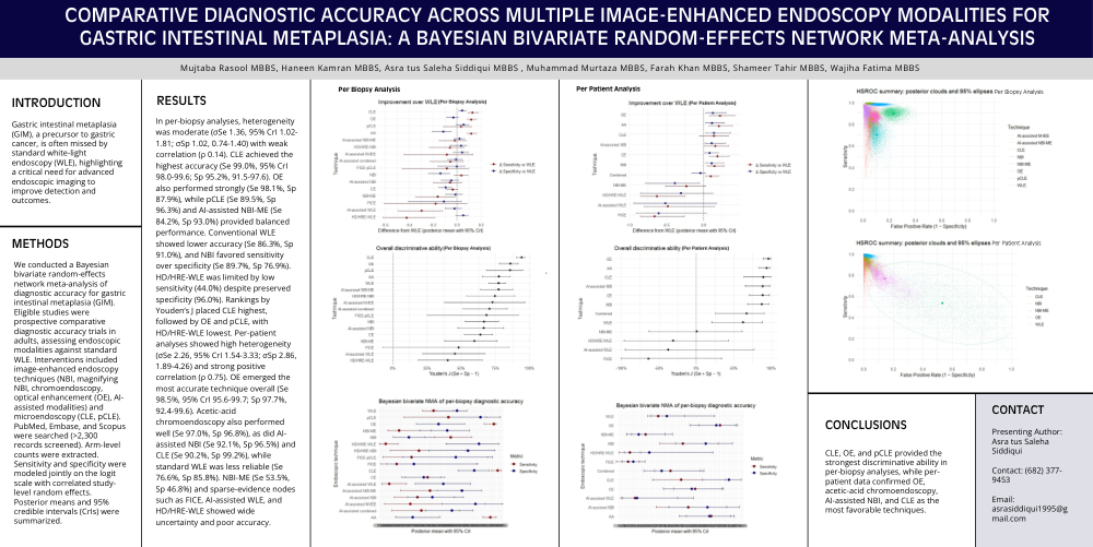
In per-biopsy analyses, heterogeneity was moderate (σSe 1.36, 95% CrI 1.02-1.81; σSp 1.02, 0.74-1.40) with weak correlation (ρ 0.14). CLE achieved the highest accuracy (Se 99.0%, 95% CrI 98.0-99.6; Sp 95.2%, 91.5-97.6). OE also performed strongly (Se 98.1%, Sp 87.9%), while pCLE (Se 89.5%, Sp 96.3%) and AI-assisted NBI-ME (Se 84.2%, Sp 93.0%) provided balanced performance. Conventional WLE showed lower accuracy (Se 86.3%, Sp 91.0%), and NBI favored sensitivity over specificity (Se 89.7%, Sp 76.9%). HD/HRE-WLE was limited by low sensitivity (44.0%) despite preserved specificity (96.0%). Rankings by Youden’s J placed CLE highest, followed by OE and pCLE, with HD/HRE-WLE lowest. Per-patient analyses showed high heterogeneity (σSe 2.26, 95% CrI 1.54-3.33; σSp 2.86, 1.89-4.26) and strong positive correlation (ρ 0.75). OE emerged the most accurate technique overall (Se 98.5%, 95% CrI 95.6-99.7; Sp 97.7%, 92.4-99.6). Acetic-acid chromoendoscopy also performed well (Se 97.0%, Sp 96.8%), as did AI-assisted NBI (Se 92.1%, Sp 96.5%) and CLE (Se 90.2%, Sp 99.2%), while standard WLE was less reliable (Se 76.6%, Sp 85.8%). NBI-ME (Se 53.5%, Sp 46.8%) and sparse-evidence nodes such as FICE, AI-assisted WLE, and HD/HRE-WLE showed wide uncertainty and poor accuracy.
Conclusion.
CLE, OE, and pCLE provided the strongest discriminative ability in per-biopsy analyses, while per-patient data confirmed OE, acetic-acid chromoendoscopy, AI-assisted NBI, and CLE as the most favorable techniques.