875 posters, 25 topics, 3,440 authors, 1,061 institutions
ePostersLive by SciGen Technologies S.A. All rights reserved.
SAGES 2026 Annual Meeting Emerging Technology Session
March 25-28, 2026 | Tampa, FL, USA

P443
Incidental Discovery of Colon Perforation by Ventriculoperitoneal Shunt Migration Requiring Sigmoidectomy
Poster Presenter
Part of Topic
Colorectal
INCIDENTAL DISCOVERY OF COLON PERFORATION BY VENTRICULOPERITONEAL SHUNT MIGRATION REQUIRING SIGMOIDECTOMYJeffrey Baum, MD1; Paige Varney, MD1; Joe Caliendo, BS2; Hsu-Tsai Chi, MD, FACS1; 1Icahn School of Medicine at Mount Sinai Hospital, Mount Sinai South Nassau; 2University of Miami
Background: Ventriculoperitoneal shunts (VPS) are widely used for hydrocephalus but carry risks of infection, malfunction, and catheter tip migration. Among the rarest complications is bowel perforation, reported in 0.01–0.07% of cases, most often involving the sigmoid colon or rectum.[1],[2] If unrecognized, this can result in peritonitis, meningitis, or death.[3] Notably, patients can remain asymptomatic, which complicates a timely diagnosis.
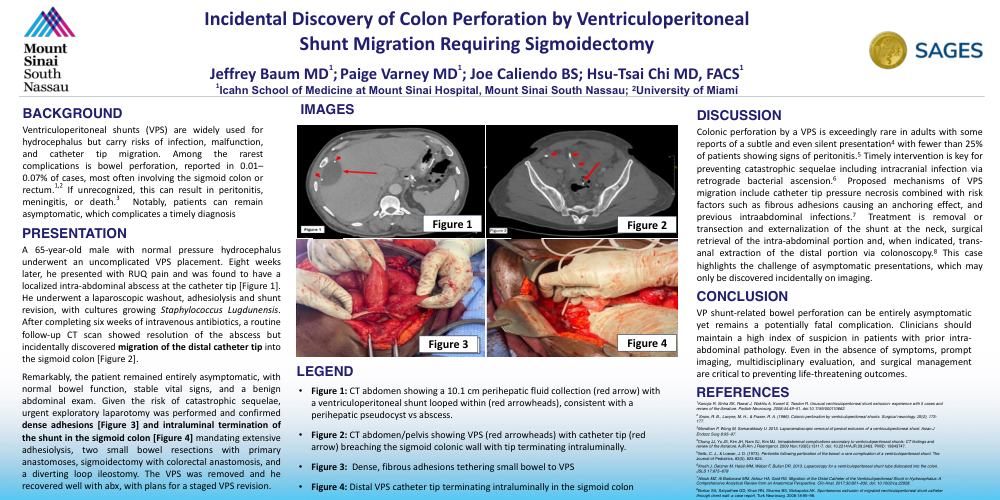
Case Presentation: A 65-year-old male with normal pressure hydrocephalus underwent an uncomplicated VPS placement. Eight weeks later, he presented with right upper quadrant pain and was found to have a localized intra-abdominal abscess at the catheter tip [Figure 1]. He underwent a laparoscopic washout, adhesiolysis and shunt revision, with cultures growing Staphylococcus lugdunensis. After completing six weeks of intravenous antibiotics, a routine follow-up CT scan showed resolution of the abscess but incidentally discovered migration of the distal catheter tip into the sigmoid colon [Figure 2].
Remarkably, the patient remained entirely asymptomatic, with normal bowel function, stable vital signs, and a benign abdominal exam. Given the risk of catastrophic sequelae, urgent exploratory laparotomy was performed and confirmed dense adhesions and intraluminal termination of the shunt in the sigmoid colon [Figures 3 and 4]. Operative interventions included extensive adhesiolysis, two small bowel resections with primary anastomoses, sigmoidectomy with colorectal anastomosis, and a diverting loop ileostomy. The VPS was removed entirely. He recovered well with antibiotics and was discharged home with plans for a staged VPS revision.
Discussion: Colonic perforation by a VPS is exceedingly rare in adults with some reports of a subtle and even silent presentation,[4] with fewer than 25% of patients showing signs of peritonitis.[5] Timely intervention is key for preventing peritonitis and intracranial infection via retrograde bacterial ascension.[6] Proposed mechanisms of VPS migration include catheter tip pressure necrosis combined with risk factors such as fibrous adhesions causing an anchoring effect, and previous intraabdominal infections.[7] Treatment is transection and externalization of the shunt at the neck, surgical retrieval of the intra-abdominal portion and, when indicated, trans-anal extraction of the distal portion via colonoscopy.[8] This case highlights the challenge of asymptomatic presentations, which may only be discovered incidentally on imaging.
Conclusion: VP shunt-related bowel perforation can be entirely asymptomatic yet remains a potentially fatal complication. Clinicians should maintain a high index of suspicion in patients with prior intra-abdominal pathology. Even in the absence of symptoms, prompt imaging, multidisciplinary evaluation, and surgical management are critical to preventing life-threatening outcomes.