875 posters, 25 topics, 3,440 authors, 1,061 institutions
ePostersLive by SciGen Technologies S.A. All rights reserved.
SAGES 2026 Annual Meeting Emerging Technology Session
March 25-28, 2026 | Tampa, FL, USA

P166
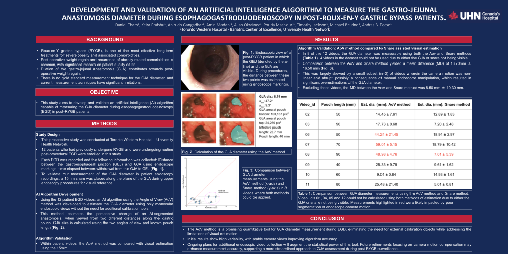
Development and validation of an artificial intelligence algorithm to measure the gastro-jejunal anastomosis diameter during esophagogastroduodenoscopy in post-Roux-en-Y gastric bypass patients.
Poster Presenter
Part of Topic
Artificial Intelligence
DEVELOPMENT AND VALIDATION OF AN ARTIFICIAL INTELLIGENCE ALGORITHM TO MEASURE THE GASTRO-JEJUNAL ANASTOMOSIS DIAMETER DURING ESOPHAGOGASTRODUODENOSCOPY IN POST-ROUX-EN-Y GASTRIC BYPASS PATIENTS.
Daniel Tham1, Keira Prabhu1, Anirudh Gangadhar1, Amin Madani1, Allan Okrainec1, Pouria Mashouri1, Timothy Jackson1, Michael Brudno1, Andras B. Fecso1.
1. Toronto Western Hospital -Bariatric Center of Excellence, University Health Network
BACKGROUND
Roux-en-Y gastric bypass (RYGB), is one of the most effective long-term treatments for severe obesity and associated comorbidities.
Post-operative weight regain and recurrence of obesity-related comorbidities is common, with significant impacts on patient quality of life.
Dilation of the gastro-jejunal anastomosis (GJA) contributes towards post-operative weight regain.
There is no gold standard measurement technique for the GJA diameter, and current measurement techniques have significant limitations.
OBJECTIVE
This study aims to develop and validate an artificial intelligence (AI) algorithm capable of measuring the GJA diameter during esophagogastroduodenoscopy (EGD) in post-RYGB patients.
METHODS
Study Design
This prospective study was conducted at Toronto Western Hospital – University Health Network.
12 patients who had previously undergone RYGB and were undergoing routine post-procedural EGD were enrolled in this study.
Each EGD was recorded and the following information was collected: Distance between the gastroesophageal junction (GEJ) and GJA using endoscope markings, time elapsed between withdrawal from the GJA to GEJ (Fig. 1).
To validate our measurement of the GJA diameter in patient endoscopy recordings, a 15mm snare was placed along the plane of the GJA during upper endoscopy procedures for visual reference.
AI Algorithm Development
Using the 12 patient EGD videos, an AI algorithm using the Angle of View (AoV) method was developed to estimate the GJA diameter using only monocular endoscopic views without the need for additional calibration tools.
This method estimates the perspective change of an AI-segmented anastomosis, when viewed from two different distances along the gastric pouch. GJA size is calculated using the two angles of view and known pouch length (Fig. 2).
Algorithm Validation
Within patient videos, the AoV method was compared with visual estimation using the 15mm.
RESULTS
Algorithm Validation: AoV method compared to Snare assisted visual estimation
In 8 of the 12 videos, the GJA diameter was measurable using both the Aov and Snare methods (Table 1). 4 videos in the dataset could not be used due to either the GJA or snare not being visible.
Comparison between the AoV and Snare method yielded a mean difference (MD) of 18.70mm ± 16.50 mm (Fig. 3).
This was largely skewed by a small subset (n=3) of videos wherein the camera motion was non-linear and abrupt, possibly a consequence of manual endoscope manipulation, which resulted in significant overestimations of the GJA diameter.
Excluding these videos, the MD between the AoV and Snare method was 8.50 mm ± 10.30 mm.
CONCLUSION
The AoV method is a promising quantitative tool for GJA diameter measurement during EGD, eliminating the need for external calibration objects while addressing the limitations of visual estimation.
Initial results show high variability, with stable camera views improving algorithm accuracy.
Ongoing plans for additional endoscopic video collection will augment the statistical power of this tool. Future refinements focusing on camera motion compensation may enhance measurement accuracy, supporting a more streamlined approach to GJA assessment during post-RYGB surveillance.
FIGURES AND TABLES
Fig. 1: Endoscopic view of a post-RYGB patient in which the GEJ (denoted by the z-line) and the GJA are visible. During procedures, the distance between these two points was estimated using endoscope markings.
Fig. 2: Calculation of the GJA diameter using the AoV method
Fig. 3: Comparison between GJA diameter measurements using the AoV method (x-axis) and Snare method (y-axis) in 8 videos where both methods could be applied.
Table 1: Comparison between GJA diameter measurements using the AoV method and Snare method. Video_id’s 01, 04, 05 and 12 could not be calculated using both methods of estimation due to either the GJA or snare not being visible. Measurements highlighted in red were likely impacted by poor segmentation or endoscope camera motion.