119 posters, 6 topics, 524 authors, 243 institutions
ePostersLive by SciGen Technologies S.A. All rights reserved.
RCR Global AI Conference 2026
29-30 June, 2026 | QEII Centre, Westminster

40
<p>Automated Uterine Fibroid Mapping on MRI Using Deep Learning: Development and Proof of Concept</p>
Poster Presenter
Part of Topic
AI Education and research: examples of proof of concept or AI in development, technical advances, teaching approaches or pre-clinical testing
AUTOMATED UTERINE FIBROID MAPPING ON MRI USING DEEP LEARNING: DEVELOPMENT AND PROOF OF CONCEPT
Meghana Menon¹, Amala Sunder², Hani Al Fadhel², Basma Darwish²
¹ Hull University Teaching Hospitals NHS Trust, UK ² Bahrain Defense Force, Royal Medical Services, Bahrain

BACKGROUND & RATIONALE
• Uterine fibroids are among the most common gynaecological conditions.
• MRI offers excellent anatomical detail, but manual fibroid delineation is time-intensive and operator-dependent.
• Inter-reader variability can affect lesion localisation, volume assessment, treatment stratification, surgical planning, and reporting consistency.
• Deep learning offers an opportunity to automate and standardise fibroid mapping.

AIM
To develop and evaluate an AI-powered deep-learning pipeline for automated uterine fibroid segmentation on MRI, as a proof-of-concept foundation for reproducible clinical decision support.

METHODS
Retrospective single-centre dataset of 98 women with confirmed uterine fibroids.
MRI performed on 1.5T and 3T scanners using a multiparametric pelvic protocol.
Two independent radiologists annotated anonymised datasets primarily on T2-weighted images in axial, sagittal, and coronal planes.
T1-weighted, diffusion-weighted, and post-contrast sequences were used where available for tissue characterisation and exclusion of alternative pathology.
A U-Net encoder–decoder convolutional neural network was trained using a 75% training / 50% validation / 50% testing split.
Structured preprocessing and augmentation were used.
Performance metrics: Dice similarity coefficient (DSC), sensitivity, specificity, precision, and F1-score.

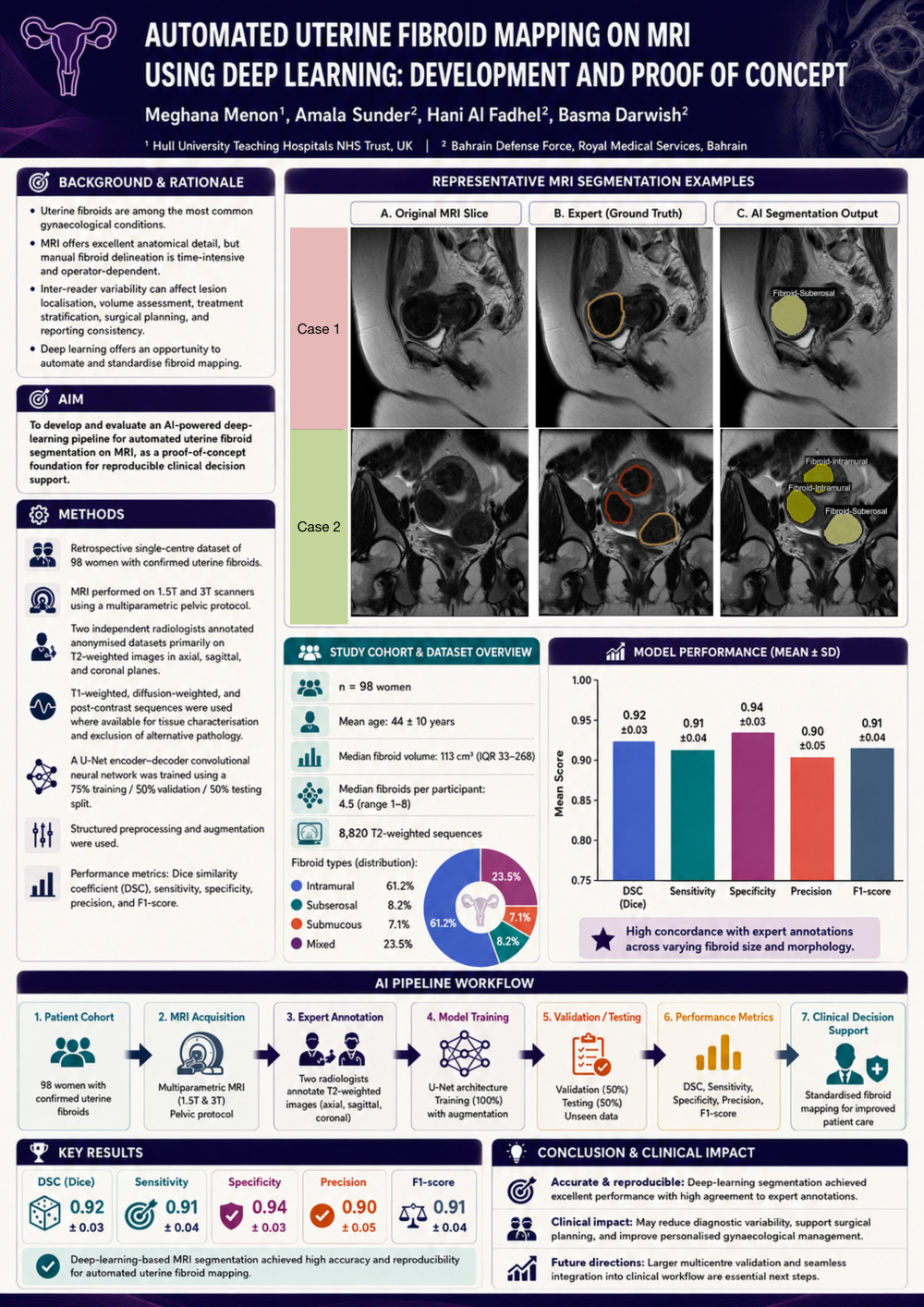
REPRESENTATIVE MRI SEGMENTATION EXAMPLES
A. Original MRI Slice B. Expert (Ground Truth) C. AI Segmentation Output
Case 1 Case 2

AI segmentation demonstrates strong concordance with expert annotations across fibroid types and locations.

STUDY COHORT & DATASET OVERVIEW
n = 98 women
Mean age: 44 ± 10 years
Median fibroid volume: 113 cm³ (IQR 33–268)
Median fibroids per participant: 4.5 (range 1–8)
8,820 T2-weighted sequences
Fibroid types (distribution): Intramural 61.2% Subserosal 8.2% Submucous 7.1% Mixed 23.5%

MODEL PERFORMANCE (MEAN ± SD)
DSC (Dice): 0.92 ± 0.03
Sensitivity: 0.91 ± 0.04
Specificity: 0.94 ± 0.03
Precision: 0.90 ± 0.05
F1-score: 0.91 ± 0.04

High concordance with expert annotations across varying fibroid size and morphology.

AI PIPELINE WORKFLOW
1. Patient Cohort 98 women with confirmed uterine fibroids
2. MRI Acquisition Multiparametric MRI (1.5T & 3T) Pelvic protocol
3. Expert Annotation Two radiologists annotate T2-weighted images (axial, sagittal, coronal)
4. Model Training U-Net architecture Training (100%) with augmentation
5. Validation / Testing Validation (50%) Testing (50%) Unseen data
6. Performance Metrics DSC, Sensitivity, Specificity, Precision, F1-score
7. Clinical Decision Support Standardised fibroid mapping for improved patient care

KEY RESULTS
DSC (Dice) 0.92 ± 0.03
Sensitivity 0.91 ± 0.04
Specificity 0.94 ± 0.03
Precision 0.90 ± 0.05
F1-score 0.91 ± 0.04

Deep-learning-based MRI segmentation achieved high accuracy and reproducibility for automated uterine fibroid mapping.

CONCLUSION & CLINICAL IMPACT
Accurate & reproducible: Deep-learning segmentation achieved excellent performance with high agreement to expert annotations.
Clinical impact: May reduce diagnostic variability, support surgical planning, and improve personalised gynaecological management.
Future directions: Larger multicentre validation and seamless integration into clinical workflow are essential next steps.