296 posters, 7 videos, 13 audios, 14 topics, 10 sessions, 1,019 authors, 260 institutions
ePostersLive by SciGen Technologies S.A. All rights reserved.
RCOphth Annual Congress 2026
18 - 21 May, 2026 | Manchester Central, Manchester

P256
Dural arteriovenous fistula in a child presenting with decompensating esophoria
Poster Presenter
Part of Topic
Paediatric ophthalmology
Background
• Intracranial dural arteriovenous fistulas (DAVFs) are abnormal shunts between dural arteries and dural venous sinuses.
• Paediatric DAVFs are rare.
• Previous reports of association with developmental delay or congestive cardiac failure.
.
Objectives & Methods
• Case report of a child with no medical history of note presenting with decompensating esophoria
• Interesting clinical presentation with subsequent investigations revealing a DAVF
Initial presentation
• 7-year-old boy
• Presented to Eye Casualty with horizontal diplopia
• No headache, nausea or vomiting
• Orthoptic examination: no abduction deficit, 30PD base
out left esotropia
• Refraction:
• Right eye: 0/-0.25 x150
• Left eye: +0.25/-0.50 x 70
2 months later
• Worsening esotropia
• Blood investigations: autoimmune or inflammatory
screen negative
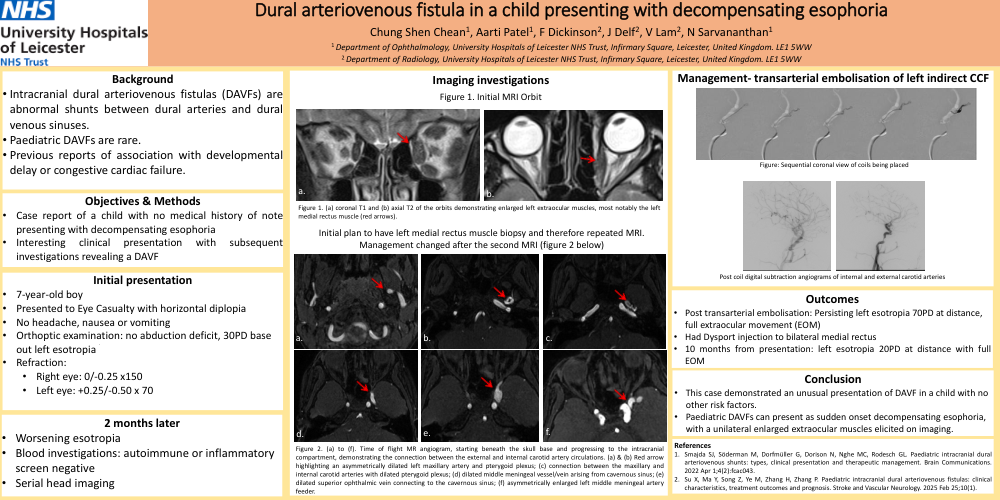
• Serial head imaging
Outcomes
• Post transarterial embolisation: Persisting left esotropia 70PD at distance, full extraocular movement (EOM)
• Had Dysport injection to bilateral medial rectus
• 10 months from presentation: left esotropia 20PD at distance with full EOM
Conclusion
• This case demonstrated an unusual presentation of DAVF in a child with no other risk factors.
• Paediatric DAVFs can present as sudden onset decompensating esophoria, with a unilateral enlarged extraocular muscles elicited on imaging.