296 posters, 7 videos, 13 audios, 14 topics, 10 sessions, 1,019 authors, 260 institutions
ePostersLive by SciGen Technologies S.A. All rights reserved.
RCOphth Annual Congress 2026
18 - 21 May, 2026 | Manchester Central, Manchester

P109
Evaluation of Macular Vessels Density Changes in Patients with Primary Open Angle Glaucoma by Swept-source Optical Coherence Tomography Angiography
Poster Presenter
Part of Topic
Glaucoma
Evaluation of Macular Vessels Density Changes in Patients with Primary Open Angle Glaucoma by Swept-source Optical Coherence Tomography Angiography
Mohamed M. Almbsut, Amira G. Abdelhameed, Dalia Sabry, Asaad A. Ghanem
Mansoura Ophthalmic Center, Faculty of Medicine, Mansoura University
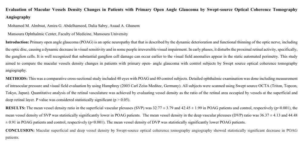
Introduction: Primary open angle glaucoma (POAG) is an optic neuropathy that that is described by the dynamic deterioration and functional thinning of the optic nerve, including the optic disc, causing a dynamic decrease in visual sensitivity and in some people irreversible visual impairment. In early phases, it disturbs the proximal retinal activity, specifically, the ganglion cells. It is well recognized that substantial ganglion cell damage can occur earlier to the visual field anomalies appear in the static automated perimetry. This study aimed to compare the macular vessels density changes in patients with primary open- angle glaucoma with control subjects by Swept source optical coherence tomography angiography.
METHODS: This was a comparative cross-sectional study included 40 eyes with POAG and 40 control subjects. Detailed ophthalmic examination was done including measurement of intraocular pressure and visual field evaluation by using Humphrey (2003 Carl Zeiss Meditec, Germany). All subjects were scanned using Swept source OCTA (Triton, Topcon, Tokyo, Japan). Quantitative analysis of the retinal vasculature was achieved by evaluating vessel density as the ratio of the retinal area occupied by vessels at the superficial and deep retinal layer. P value was considered statistically significant (p > 0.05).
RESULTS: The mean vessel density ratio in the superficial vascular plexuses (SVP) was 32.77 ± 3.79 and 42.45 ± 1.99 in POAG patients and control, respectively (p<0.001), the mean vessel density of SVP was statistically significantly lower in POAG patients. The mean vessel density in the deep vascular plexuses (DVP) ratio was 36.37 ± 4.13 and 44.48 ± 0.91 in POAG patients and control, respectively (p<0.001). The mean vessel density of DVP was statistically significantly lower POAG patients.
CONCLUSION: Macular superficial and deep vessel density by Swept-source optical coherence tomography angiography showed statistically significant decrease in POAG patients.