197 posters, 15 videos, 1 topics, 31 sessions, 509 authors
ePostersLive by SciGen Technologies S.A. All rights reserved.
2026 AO Annual Meeting
Conference ePosters

P210
Comparison of Soft Tissue Dimensional Changes Between Two Different Suturing Techniques Using the VISTA Technique: In-Vitro Pig Jaw Model
Poster Presenter
Authors
Part of Topic
Comparison of soft tissue dimensional changes between two different suturing techniques using the VISTA technique: in-vitro pig jaw model
Liza Harutyunyan, DDS, Homayoun H. Zadeh, DDS, PhD, Yusuke Hamada, DDS, MSD
Introduction:
The vestibular incision subperiosteal tunnel access (VISTA) technique has become increasingly adopted for phenotype modification and root coverage in periodontal and peri-implant soft tissue therapy.
The technique involves a remote vestibular access incision, subperiosteal tunnel preparation, coronal advancement of the gingival margin, and stabilization using sutures bonded to the mid-coronal tooth surface.
Despite its widespread use, the influence of different suturing strategies on coronal tissue advancement and soft-tissue thickness has not been fully elucidated.
Aim:
The vestibular incision subperiosteal tunnel access (VISTA) technique has become increasingly adopted for phenotype modification and root coverage in periodontal and peri-implant soft tissue therapy.
The technique involves a remote vestibular access incision, subperiosteal tunnel preparation, coronal advancement of the gingival margin, and stabilization using sutures bonded to the mid-coronal tooth surface.
Despite its widespread use, the influence of different suturing strategies on coronal tissue advancement and soft-tissue thickness has not been fully elucidated.
Methods:
One surgeon performed the procedures using VISTA on the right and left sides of the mandibular third and fourth deciduous diphyodonts (DDP3, DDP4), and deciduous first and second molars (M1, M2) in four pig jaw models.
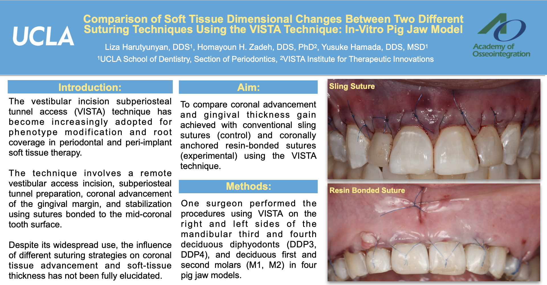
On one side of the porcine mandible, coronal stabilization of the tunneled gingival tissues was achieved using a double mattress suturing technique. A monofilament suture (6.0 polypropylene with C3 needle) was inserted at the base of the mesial papilla and passed through the tunnel to emerge approximately 5 mm apical to the mesial line angle of the experimental tooth. At the same apico-coronal level, the needle was reinserted at the distal line angle and advanced through the tunnel to exit at the base of the distal papilla.
A loop of suture was maintained in the apical portion of the tunnel to allow passive coronal advancement of the tissues. The gingival margin was positioned coronally with the aid of an instrument placed within the tunnel, and the suture was tied using a double knot without excessive tension. The suture knot was stabilized to the facial enamel surface by spot etching, followed by application and light polymerization of flowable composite resin to maintain coronal tissue position.
On the contralateral side of the mandible, coronal stabilization was performed using a sling suture technique and served as the control procedure.
Following tunnel preparation, a monofilament suture was introduced at the base of the mesial papilla and passed through the tunnel to emerge approximately 5 mm apical to the mesial line angle.
The suture was then passed through the mesial embrasure to the lingual aspect, wrapped circumferentially around the cervical portion of the tooth, and returned to the buccal aspect through the distal embrasure. The needle was reintroduced on the distal aspect at the same apico-coronal level and passed through the tunnel to complete the sling configuration.
Gentle tension was applied to advance the gingival margin coronally, and the suture was secured on the facial aspect with a square knot.
Pre- and post-operative surface-scanner models were obtained as Standard Tessellation Language (STL) files and superimposed to measure soft-tissue thickness changes and the distance of coronal advancement in both groups.
Post-operative gingival margin (GM) was used as a reference.
Soft tissue thickness changes were measured at 1-5mm apical to the gingival margin (GM1mm - GM5mm) at the facial side of each cusp of the treated teeth.
The coronal advancement was measured on the facial side of each cusp area as well. Measurements were performed on two cusps of DDP3, and three cusps of DDP4 and M1.
Results:
The coronal advancement was 0.73 ± 0.28 mm in the control group, and 3.42 ± 0.99 mm in the test group, and the difference was statistically significant (p<0.05).
In the control group, soft tissue thickness changes were 1.04 ± 0.39 mm, 1.09 ± 0.49 mm, 1.11 ± 0.48mm, 1.03 ± 0.54mm, and 1.01 ± 0.59mm from GM1mm to GM5 mm, respectively.
In the test group, the changes were 1.97 ± 0.73 mm, 2.16 ± 0.81 mm, 2.23 ± 0.82 mm, 2.48 ± 0.83 mm, and 2.64 ± 0.81 mm from GM1 mm to GM5 mm, respectively.
The changes in the test group were significantly higher than in the control group (p<0.05)
Conclusion:
Within the limitations of this study, coronally anchored suturing demonstrated greater coronal advancement and increased soft-tissue thickness compared with the sling suture technique.
Resin-bonded suture stabilization may therefore enhance phenotype modification therapy, which is increasingly regarded as a primary therapeutic objective in the management of gingival recession defects.