301 posters, 50 videos, 13 topics, 13 sessions, 734 authors, 193 institutions
ePostersLive by SciGen Technologies S.A. All rights reserved.
ISMICS 2026 Annual Meeting
10 - 13 June, 2026 | Miami, Florida

P79
Micro-perfusion Concept For All Minimally Invasive And Re-do Procedures With True Antegrade Perfusion - A Case Series
Poster Presenter
Part of Topic
Heart Failure Assist Devices, Heart Transplantation & Perfusion
Funding Statement: The Austrian Federal Ministry of Labour and Economy, the National Foundation for Research, Technology, and Development, and the Christian Doppler Research Association funded part of the research.
OBJECTIVE
Peripheral femoral cannulation is common in µ-invasive cardiac surgery, but retrograde aortic perfusion may increase stroke risk in atherosclerotic patients, thus potentially limiting its use in aortic stenosis or coronary disease. We standardized our percutaneous technique with a long arterial cannula to enable antegrade perfusion in almost all minimally invasive or redo cases.
METHODS
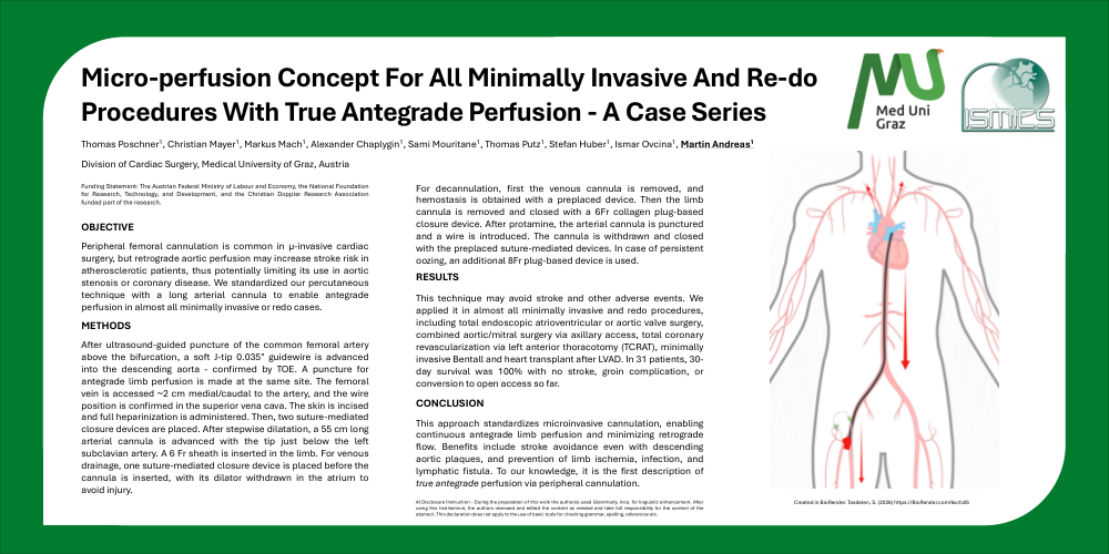
After ultrasound-guided puncture of the common femoral artery above the bifurcation, a soft J-tip 0.035″ guidewire is advanced into the descending aorta - confirmed by TOE. A puncture for antegrade limb perfusion is made at the same site. The femoral vein is accessed ~2 cm medial/caudal to the artery, and the wire position is confirmed in the superior vena cava. The skin is incised and full heparinization is administered. Then, two suture-mediated closure devices are placed. After stepwise dilatation, a 55 cm long arterial cannula is advanced with the tip just below the left subclavian artery. A 6 Fr sheath is inserted in the limb. For venous drainage, one suture-mediated closure device is placed before the cannula is inserted, with its dilator withdrawn in the atrium to avoid injury.
For decannulation, first the venous cannula is removed, and hemostasis is obtained with a preplaced device. Then the limb cannula is removed and closed with a 6Fr collagen plug-based closure device. After protamine, the arterial cannula is punctured and a wire is introduced. The cannula is withdrawn and closed with the preplaced suture-mediated devices. In case of persistent oozing, an additional 8Fr plug-based device is used.
RESULTS
This technique may avoid stroke and other adverse events. We applied it in almost all minimally invasive and redo procedures, including total endoscopic atrioventricular or aortic valve surgery, combined aortic/mitral surgery via axillary access, total coronary revascularization via left anterior thoracotomy (TCRAT), minimally invasive Bentall and heart transplant after LVAD. In 31 patients, 30- day survival was 100% with no stroke, groin complication, or conversion to open access so far.
CONCLUSION
This approach standardizes microinvasive cannulation, enabling continuous antegrade limb perfusion and minimizing retrograde flow. Benefits include stroke avoidance even with descending aortic plaques, and prevention of limb ischemia, infection, and lymphatic fistula. To our knowledge, it is the first description of true antegrade perfusion via peripheral cannulation.