301 posters, 50 videos, 13 topics, 13 sessions, 734 authors, 193 institutions
ePostersLive by SciGen Technologies S.A. All rights reserved.
ISMICS 2026 Annual Meeting
10 - 13 June, 2026 | Miami, Florida

D83
Preserving Breastfeeding And Cosmesis: Robotic Aortic Valve Tumor Resection In A Young Mother
Poster Presenter
Part of Topic
Aortic and Endovascular Therapies
OBJECTIVE: Papillary fibroelastomas (PFE) are rare benign cardiac tumors commonly arising on valvular surfaces and are associated with embolic events. Surgical resection is recommended in symptomatic patients or when tumors are greater than 1 cm. While robotic platforms are increasingly utilized in mitral valve and atrial mass resections, its application in aortic valve (AV) tumors remains limited due to anatomical and technical challenges. We report the robotic resection of a 1.6 cm PFE originating from the left coronary cusp in an actively breastfeeding mother, with a focus on surgical adaptation to her postpartum physiology and lactation status.
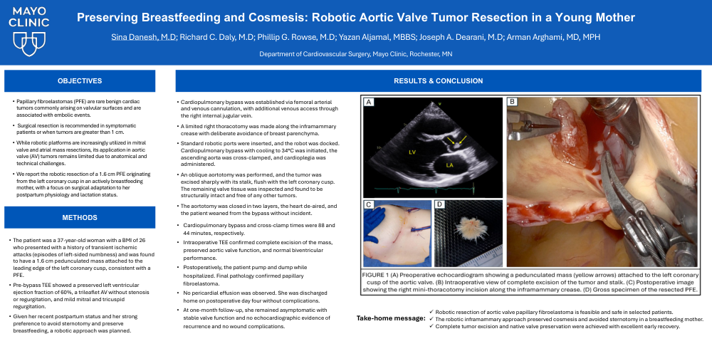
METHODS: The patient was a 37-year-old woman with a BMI of 26 who presented with a history of transient ischemic attacks (episodes of left-sided numbness) and was found to have a 1.6 cm pedunculated mass attached to the leading edge of the left coronary cusp, consistent with a PFE. Pre-bypass TEE showed a preserved left ventricular ejection fraction of 60%, a trileaflet AV without stenosis or regurgitation, and mild mitral and tricuspid regurgitation. Given her recent postpartum status and her strong preference to avoid sternotomy and preserve breastfeeding, a robotic approach was planned. Cardiopulmonary bypass was established via femoral arterial and venous cannulation, with additional venous access through the right internal jugular vein. A limited right thoracotomy was made along the inframammary crease with deliberate avoidance of breast parenchyma. Standard robotic ports were inserted, and the robot was docked. Cardiopulmonary bypass with cooling to 34°C was initiated, the ascending aorta was cross-clamped, and cardioplegia was administered. An oblique aortotomy was performed, and the tumor was excised sharply with its stalk, flush with the left coronary cusp. The remaining valve tissue was inspected and found to be structurally intact and free of any other tumors. The aortotomy was closed in two layers, the heart de-aired, and the patient weaned from the bypass without incident. (Figure 1)
RESULTS: Cardiopulmonary bypass and cross-clamp times were 88 and 44 minutes, respectively. Intraoperative TEE confirmed complete excision of the mass, preserved aortic valve function, and normal biventricular performance. Postoperatively, the patient pump and dump while hospitalized. Final pathology confirmed papillary fibroelastoma. No pericardial effusion was observed. She was discharged home on postoperative day four without complications. At one-month follow-up, she remained asymptomatic with stable valve function and no echocardiographic evidence of recurrence and no wound complications.
CONCLUSIONS: Robotic resection of aortic PFE is a viable and safe option in selected patients. In this postpartum, actively breastfeeding patient, a tailored minimally invasive approach enabled complete tumor excision while minimizing disruption to lactation and preserving both cosmesis and functional recovery. This case underscores the evolving role of robotic platforms in managing complex valvular pathology.