301 posters, 50 videos, 13 topics, 13 sessions, 734 authors, 193 institutions
ePostersLive by SciGen Technologies S.A. All rights reserved.
ISMICS 2026 Annual Meeting
10 - 13 June, 2026 | Miami, Florida

P145
Tavr Explantation Via Right Anterior Minithoracotomy - Tips And Special Considerations
Poster Presenter
Authors
Part of Topic
Aortic and Endovascular Therapies
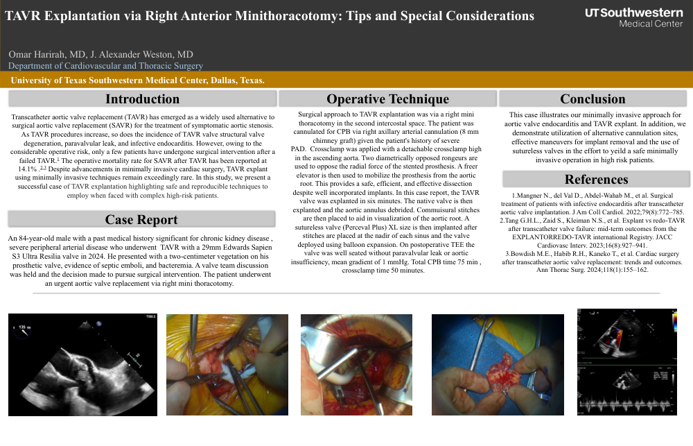
Introduction Transcatheter aortic valve replacement (TAVR) has emerged as a widely used alternative to Operative Technique Surgical approach to TAVR explantation was via a right mini Conclusion surgical aortic valve replacement (SAVR) for the treatment of symptomatic aortic stenosis. As TAVR procedures increase, so does the incidence of TAVR valve structural valve degeneration, paravalvular leak, and infective endocarditis. However, owing to the considerable operative risk, only a few patients have undergone surgical intervention after a failed TAVR.1 The operative mortality rate for SAVR after TAVR has been reported at 14.1% .2 ,3 Despite advancements in minimally invasive cardiac surgery, TAVR explant using minimally invasive techniques remain exceedingly rare. In this study, we present a successful case of TAVR explantation highlighting safe and reproducible techniques to employ when faced with complex high-risk patients. Case Report An 84-year-old male with a past medical history significant for chronic kidney disease , severe peripheral arterial disease who underwent TAVR with a 29mm Edwards Sapien S3 Ultra Resilia valve in 2024. He presented with a two-centimeter vegetation on his prosthetic valve, evidence of septic emboli, and bacteremia. A valve team discussion was held and the decision made to pursue surgical intervention. The patient underwent an urgent aortic valve replacement via right mini thoracotomy. thoracotomy in the second intercostal space. The patient was cannulated for CPB via right axillary arterial cannulation (8 mm chimney graft) given the patient's history of severe PAD. Crossclamp was applied with a detachable crossclamp high in the ascending aorta. Two diametrically opposed rongeurs are used to oppose the radial force of the stented prosthesis. A freer elevator is then used to mobilize the prosthesis from the aortic root. This provides a safe, efficient, and effective dissection despite well incorporated implants. In this case report, the TAVR valve was explanted in six minutes. The native valve is then explanted and the aortic annulus debrided. Commuisural stitches are then placed to aid in visualization of the aortic root. A sutureless valve (Perceval Plus) XL size is then implanted after stitches are placed at the nadir of each sinus and the valve deployed using balloon expansion. On postoperative TEE the valve was well seated without paravalvular leak or aortic insufficiency, mean gradient of 1 mmHg. Total CPB time 75 min , crossclamp time 50 minutes. This case illustrates our minimally invasive approach for aortic valve endocarditis and TAVR explant. In addition, we demonstrate utilization of alternative cannulation sites, effective maneuvers for implant removal and the use of sutureless valves in the effort to yeild a safe minimally invasive operation in high risk patients. References 1.Mangner N., del Val D., Abdel-Wahab M., et al. Surgical treatment of patients with infective endocarditis after transcatheter aortic valve implantation. J Am Coll Cardiol. 2022;79(8):772–785. 2.Tang G.H.L., Zaid S., Kleiman N.S., et al. Explant vs redo-TAVR after transcatheter valve failure: mid-term outcomes from the EXPLANTORREDO-TAVR international Registry. JACC Cardiovasc Interv. 2023;16(8):927–941. 3.Bowdish M.E., Habib R.H., Kaneko T., et al. Cardiac surgery after transcatheter aortic valve replacement: trends and outcomes. Ann Thorac Surg. 2024;118(1):155–162