395 posters, 1 audios, 13 topics, 29 sessions, 1,056 authors, 461 institutions
ePostersLive by SciGen Technologies S.A. All rights reserved.
51st Annual Regional Anesthesiology and Acute Pain Medicine Meeting
April 16 - 18, 2026 | Phoenix, Arizona

2313868
Preoperative gastric ultrasound assessment in patients undergoing post-traumatic fracture surgery
Poster Presenter
Authors
Affiliations
Part of Topic
Scientific Abstracts > Emerging Technology
Preoperative Gastric Ultrasound Assessment in Post-Traumatic Fracture Surgery
Introduction
Pulmonary aspiration of gastric content is a potentially preventable perioperative complication associated with significant morbidity and mortality (1). Although preoperative fasting guidelines are widely applied, they may not reliably reflect actual gastric content in certain patient populations. Trauma-related inflammatory responses and increased stress hormone levels following fractures may impair gastric emptying, even in patients who report adequate fasting (2). However, data on preoperative gastric ultrasound assessment in patients undergoing post-traumatic fracture surgery is limited.
Materials and Methods
The study was prospectively registered at ClinicalTrials.gov (NCT05728242). Written informed consent was obtained from all participants. Adult patients (≥18 years) with American Society of Anesthesiologists (ASA) physical status I–III scheduled for orthopedic surgery within three days of traumatic fracture were included. Patients with conditions known to affect gastric emptying (including diabetes mellitus, gastroesophageal reflux disease, body mass index >35 kg/m², pregnancy, prior gastric surgery, preoperative opioid use, connective tissue disorders, hiatal hernia, and biliary tract disease) were excluded.
Preoperative gastric ultrasound was performed in the operating room by a single anesthesiologist experienced in perioperative ultrasound. Examinations were conducted in the supine and right lateral decubitus positions. Qualitative gastric content was assessed using the Perlas grading system based solely on sonographic visualization. Quantitative gastric volume was estimated in milliliters using a validated formula and classified according to quantitative Perlas grading. Weight-adjusted gastric volume was calculated separately by dividing estimated gastric volume by body weight. A high-risk stomach was defined exclusively as a weight-adjusted gastric volume >1.5 mL/kg. Qualitative, quantitative, and weight-adjusted assessments were analyzed as distinct measures. Secondary outcomes included postoperative nausea or vomiting within 24 hours and aspiration pneumonia within 48 hours.
Results
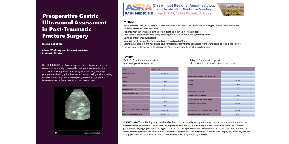
Given the preliminary nature of the study and the limited sample size (n=36), results are presented descriptively. Baseline demographic and perioperative characteristics are summarized in Table 1. Preoperative gastric ultrasound findings, including qualitative gastric content grading and quantitative risk classification, as well as postoperative clinical outcomes, are presented in Table 2 and illustrated in Figure 1. Based on quantitative Perlas classification, 20 patients (55.6%) were classified as Grade 0, 5 (13.9%) as Grade 1, and 10 patients (27.8%) as Grade 2. However, none of the patients exceeded the predefined high-risk gastric volume threshold of >1.5 mL/kg when weight-adjusted gastric volume was considered. Postoperative nausea or vomiting within the first 24 hours occurred in 10 patients (27.8%). No cases of aspiration pneumonia were observed within 48 hours postoperatively.
Discussion
This preliminary observational analysis demonstrates that preoperative gastric ultrasound is a feasible method for descriptive assessment of gastric content and aspiration risk in patients undergoing post-traumatic fracture surgery. These findings suggest that absolute volume–based grading alone may overestimate aspiration risk in post-traumatic fracture patients, highlighting the importance of weight-adjusted gastric volume evaluation in perioperative risk stratification. The absence of aspiration pneumonia, even among patients identified as having increased quantitative risk, highlights the role of gastric ultrasound as a perioperative risk-stratification tool rather than a predictor of clinical events. Given the limited sample size and descriptive scope, these findings should be interpreted cautiously and regarded as hypothesis-generating for future large-scale prospective studies (3).
References
1) Van de Putte P, Perlas A. Gastric ultrasound and aspiration risk. Br J Anaesth. 2014; 113:12–22.
2) Zhang G, Huang X, Shui Y, Luo C, Zhang L. Ultrasound to guide the individual medical decision by evaluating the gastric contents and risk of aspiration: A literature review. Asian J Surg. 2020 Dec;43(12):1142-1148.
3) Kar Man Chan L. Gastric ultrasound: Enhancing preoperative risk assessment and patient safety. J Perioper Pract. 2025 Nov;35(11):501-503.
Table 1. Baseline characteristics and perioperative variables
|
Variable |
Overall (n = 36) |
|
Age, years, mean ± SD |
54.7 ± 19.2 |
|
Sex, n (%) |
|
|
Female |
18 (50.0) |
|
Male |
17 (47.2) |
|
BMI, kg/m², mean ± SD |
24.4 ± 4.8 |
|
ASA physical status, n (%) |
|
|
I |
20 (55.6) |
|
II |
14 (38.9) |
|
III |
1 (2.8) |
|
Time from fracture to surgery, days, median (IQR) |
3.0 (2.0–3.0) |
|
Fasting time, hours, median (IQR) |
10.0 (10.0–13.5) |
Data are presented as median (IQR) or number (%), as appropriate.
Table 2. Preoperative gastric ultrasound findings and clinical outcomes
|
Outcome |
Overall (n = 36) |
|
Primary outcomes (gastric ultrasound) |
|
|
Estimated gastric volume, mL, median (IQR) |
47.0 (33.0–55.0) |
|
Qualitative gastric content grade (Perlas), n (%) |
|
|
Grade 0 |
19 (52.8) |
|
Grade 1 |
5 (13.9) |
|
Grade 2 |
11 (30.6) |
|
Quantitative risk grade, n (%) |
|
|
Grade 0 |
20 (55.6) |
|
Grade 1 |
5 (13.9) |
|
Grade 2 |
10 (27.8) |
|
High-risk gastric volume (>1.5 mL/kg), n (%) |
0 (0) |
|
Secondary outcomes |
|
|
Postoperative nausea or vomiting (0–24 h), n (%) |
10 (27.8) |
|
Aspiration pneumonia (0–48 h), n (%) |
0 (0) |
Qualitative gastric content was graded according to Perlas classification, and quantitative risk grading was based on estimated gastric volume.
Figure 1. Preoperative gastric ultrasound image demonstrating antral visualization used for qualitative grading and quantitative risk assessment.