395 posters, 1 audios, 13 topics, 29 sessions, 1,056 authors, 461 institutions
ePostersLive by SciGen Technologies S.A. All rights reserved.
51st Annual Regional Anesthesiology and Acute Pain Medicine Meeting
April 16 - 18, 2026 | Phoenix, Arizona

2310358
Ultrasound-guided bilateral TAP blocks in Takayasu arteritis with Crohn’s-related aortoiliac occlusion
Poster Presenter
Part of Topic
Medically Challenging Cases
INTRODUCTION
Takayasu’s arteritis (TA) is a rare granulomatous vasculitis that causes critical aortic and branch vessel occlusion, leaving fragile abdominal wall collaterals as the sole source of lower extremity perfusion (2,3). The coexistence of TA and Crohn’s disease has been reported and is associated with a hypercoagulable state, conferring elevated thromboembolic and ischemic risk (3). Regional anesthesia in this setting is particularly challenging , requiring effective analgesia without compromising collateral blood flow.
METHODS
The patient provided informed consent for the submission of this case report. As the case report is devoid of patient-identifiable information, it is exempt from IRB review requirements per University of North Carolina policy.
CASE REPORT
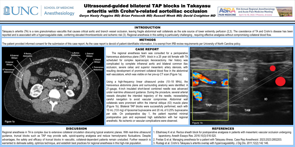
The regional anesthesia team was consulted for a perioperative transversus abdominus plane (TAP) block in a 25 year old female with TA scheduled for complex laparoscopic ileocecectomy. Her history was complicated by complete infrarenal aortic and bilateral common iliac occlusion, severe celiac and superior mesenteric artery stenosis, with resulting development of prominent collateral blood flow in the abdominal wall vasculature, which was visible on her pre-op CT scan (Figure 1a).
Using a high-frequency linear ultrasound probe (10–18 MHz), the transversus abdominis plane and surrounding anatomy were identified. A 21-gauge, 4-inch insulated short-bevel combined needle was advanced under real-time ultrasound guidance. During the procedure, several arterial vessels disrupted the intended trajectory of the needle, necessitating careful navigation to avoid vascular compromise. Abdominal wall collaterals were prominent within the internal oblique (IO) muscle plane (Figure 1b). Bilateral TAP blocks were successfully performed, each with 10 mL (133 mg) of liposomal bupivacaine and 20 mL of 0.25% bupivacaine per side. On postoperative day 1, the patient reported minimal postoperative pain and expressed high satisfaction with her regional anesthetic. No ischemic or vascular complications were observed.
DISCUSSION
Regional anesthesia in TA is complex due to extensive collateral circulation obscuring typical anatomic planes. With real-time ultrasound guidance, truncal blocks such as TAP may provide safe, opioid-sparing analgesia and reduce hemodynamic fluctuations. Despite advantages, the safety and efficacy of truncal blocks in vasculitic, collateral-dependent patients remain unstudied. Further research is warranted to delineate safety, optimize technique, and establish best practices for regional anesthesia in this high-risk population.
REFERENCES
1. Elbahrawy K et al. Rectus sheath block for postoperative analgesia in patients with mesenteric vascular occlusion undergoing laparotomy. Anesth Essays Res. 2016;10(3):516-520.
2. Freitas G et al. Managing anesthesia for a patient with Takayasu illness. Case Rep Anesthesiol. 2023;2023:2852203.
3. Rustagi et al. Crohn’s Takayasu’s arteritis overlap with hypercoagulability. J Dig Dis. 2011;12(2):142 146.