395 posters, 1 audios, 13 topics, 29 sessions, 1,056 authors, 461 institutions
ePostersLive by SciGen Technologies S.A. All rights reserved.
51st Annual Regional Anesthesiology and Acute Pain Medicine Meeting
April 16 - 18, 2026 | Phoenix, Arizona

2318751
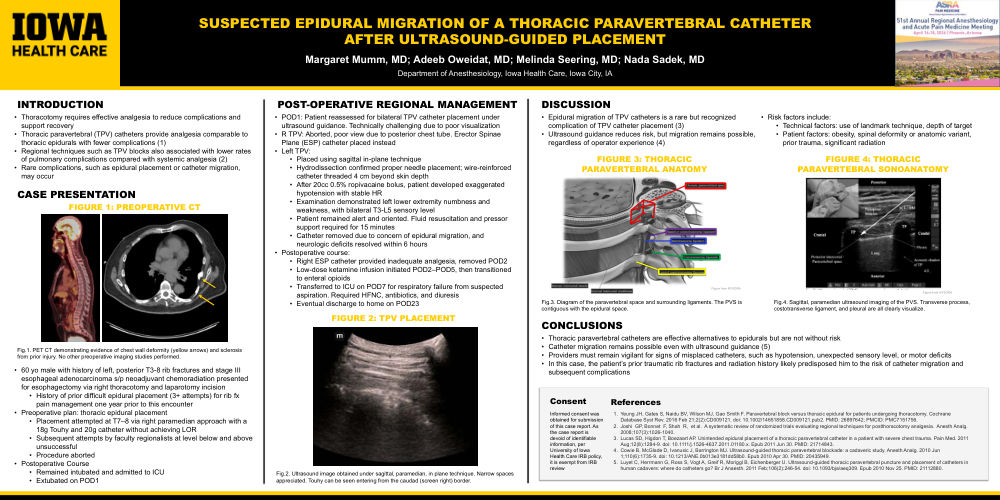
Suspected epidural migration of thoracic paravertebral catheter
Poster Presenter
Authors
Affiliations
Part of Topic
Medically Challenging Cases
INTRODUCTION
Thoracotomy requires effective analgesia to reduce complications and support recovery
Thoracic paravertebral (TPV) catheters provide analgesia comparable to thoracic epidurals with fewer complications (1)
Regional techniques such as TPV blocks also associated with lower rates of pulmonary complications compared with systemic analgesia (2)
Rare complications, such as epidural placement or catheter migration, may occur
CASE PRESENTATION
60 yo male with history of left, posterior T3-8 rib fractures and stage III esophageal adenocarcinoma s/p neoadjuvant chemoradiation presented for esophagectomy via right thoracotomy and laparotomy incision
History of prior difficult epidural placement (3+ attempts) for rib fx pain management one year prior to this encounter
Preoperative plan: thoracic epidural placement
Placement attempted at T7–8 via right paramedian approach with a 18g Touhy and 20g catheter without achieving LOR
Subsequent attempts by faculty regionalists at level below and above unsuccessful
Procedure aborted
Postoperative Course
Remained intubated and admitted to ICU
Extubated on POD1
POST-OPERATIVE REGIONAL MANAGEMENT
POD1: Patient reassessed for bilateral TPV catheter placement under ultrasound guidance. Technically challenging due to poor visualization
R TPV: Aborted, poor view due to posterior chest tube. Erector Spinae Plane (ESP) catheter placed instead
Left TPV:
Placed using sagittal in-plane technique
Hydrodissection confirmed proper needle placement; wire-reinforced catheter threaded 4 cm beyond skin depth
After 20cc 0.5% ropivacaine bolus, patient developed exaggerated hypotension with stable HR
Examination demonstrated left lower extremity numbness and weakness, with bilateral T3-L5 sensory level
Patient remained alert and oriented. Fluid resuscitation and pressor support required for 15 minutes
Catheter removed due to concern of epidural migration, and neurologic deficits resolved within 6 hours
Postoperative course:
Right ESP catheter provided inadequate analgesia, removed POD2
Low-dose ketamine infusion initiated POD2–POD5, then transitioned to enteral opioids
Transferred to ICU on POD7 for respiratory failure from suspected aspiration. Required HFNC, antibiotics, and diuresis
Eventual discharge to home on POD23
DISCUSSION
Epidural migration of TPV catheters is a rare but recognized complication of TPV catheter placement (3)
Ultrasound guidance reduces risk, but migration remains possible, regardless of operator experience (4)
Risk factors include:
Technical factors: use of landmark technique, depth of target
Patient factors: obesity, spinal deformity or anatomic variant, prior trauma, significant radiation
CONCLUSIONS
Thoracic paravertebral catheters are effective alternatives to epidurals but are not without risk
Catheter migration remains possible even with ultrasound guidance (5)
Providers must remain vigilant for signs of misplaced catheters, such as hypotension, unexpected sensory level, or motor deficits
In this case, the patient’s prior traumatic rib fractures and radiation history likely predisposed him to the risk of catheter migration and subsequent complications