395 posters, 1 audios, 13 topics, 29 sessions, 1,056 authors, 461 institutions
ePostersLive by SciGen Technologies S.A. All rights reserved.
51st Annual Regional Anesthesiology and Acute Pain Medicine Meeting
April 16 - 18, 2026 | Phoenix, Arizona

2318720
2D and 3D Anatomic Analysis of Thoracic Epidural Placement: A Single Center 1-Year Retrospective Review
Poster Presenter
Authors
Affiliations
Part of Topic
Scientific Abstracts > Regional Anesthesia
Intro:
•Success rates for thoracic epidural placement vary, and primary failure remains a concern.
•CT imaging of the spine is not routinely used for epidural planning.
•Anatomical factors may impact epidural catheter success and may be identified using 3D-rendered and quantitative 2D CT images when available.
Methods:
•1-year retrospective chart review was conducted for thoracic epidurals attempted at a single quaternary care academic medical center.
•Available CT images were systematically reviewed to evaluate anatomical factors associated with technically difficult placement
•This analysis was performed using standard 2D CT scans (three planes) and 3D-rendered images.
•A suggested best approach for epidural placement was also recorded, based on the most open space on imaging.
Results:
•There were 11 epidural placement failures among 524 total thoracic epidurals attempted (98% success rate).
•For patient characteristics, the majority were male, with varying ages and morphometrics.
•Upper abdominal surgery was the most common indication.
•The most common anatomic findings: apposition of spinous processes (< 2 mm) (n = 8, 73%); scoliosis/rotation of the spine (n = 6, 55%); and narrow interlaminar spaces (< 3 mm) (n = 5, 45%) (Table 2).
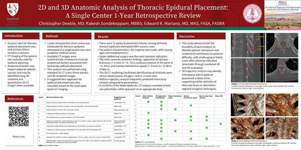
•The 3D CT renderings facilitated identification of relatively open versus closed spaces (Images 1 and 2) in each case.
•Midline trajectory analysis frequently predicted more bony contact compared to paramedian
•In 10 (91%) of the failed epidurals, CT analysis revealed at least one potentially viable approach at an appropriate level
Discussion:
•This study demonstrates the feasibility of spine analysis to identify optimal interspaces and anatomical contributors to epidural difficulty, with challenging or failed cases often showing indicators detectable through combined 2D and 3D evaluation.
•3D trajectory analysis may identify interspaces where epidural placement is likely futile, supporting earlier selection of alternate levels or alternative regional analgesic techniques.