395 posters, 1 audios, 13 topics, 29 sessions, 1,056 authors, 461 institutions
ePostersLive by SciGen Technologies S.A. All rights reserved.
51st Annual Regional Anesthesiology and Acute Pain Medicine Meeting
April 16 - 18, 2026 | Phoenix, Arizona

2308288
Inadvertent Malpositioning of Thoracic Paravertebral Catheters for Robotic Mitral Valve Repair: A Limited Case Series
Poster Presenter
Authors
Mishan Rambukwella, David Mariano, Elliot Schwartz, Jun Tang
Affiliations
Part of Topic
Medically Challenging Cases
Inadvertent Malpositioning of Thoracic Paravertebral Catheters for Robotic Mitral Valve Repair: A Limited Case Series
Mishan Rambukwella, David Mariano, Elliot Schwartz, Jun Tang
Introduction
Ultrasound-guided paravertebral blocks are effective for intraoperative and postoperative analgesia for patients undergoing mini thoracotomies. However, given the block’s anatomical parameters and patient discomfort if performed awake, this technique is also one of the most challenging regional anesthesia techniques. Here, we present two occurrences of preoperatively placed thoracic paravertebral catheters (PVCs) that were intraoperatively found to be malpositioned during robotic mitral valve repair (MVR) surgery.
Case Series
Both patients consented to have their cases used for educational and research purposes. As these cases have no unique patient identifiable information, it is exempt from Cedars-Sinai IRB review requirements as per the Determining If Activities Meet “Not Regulated” Status policy.
Case 1:
A 61M, BMI 26.3, with nonrheumatic mitral valve regurgitation presented for robotic MVR surgery via right-sided mini thoracotomy. Before induction of general anesthesia with ASA standard monitors, an ultrasound-guided T5-T6 PVC was placed. Intraoperatively, the PVC was noted to be intrapleural by the surgery team during closure. Given the intraoperative need for heparinization, the catheter was left in place. Postoperatively, the PVC was not activated, and after normalization of the patient’s INR on POD0, the malpositioned catheter was removed and successfully replaced with a new catheter under ultrasound guidance. This catheter provided appropriate analgesia at the chest tube site prior to its removal without complications on POD4 after the patient’s chest tube was removed.
Case 2:
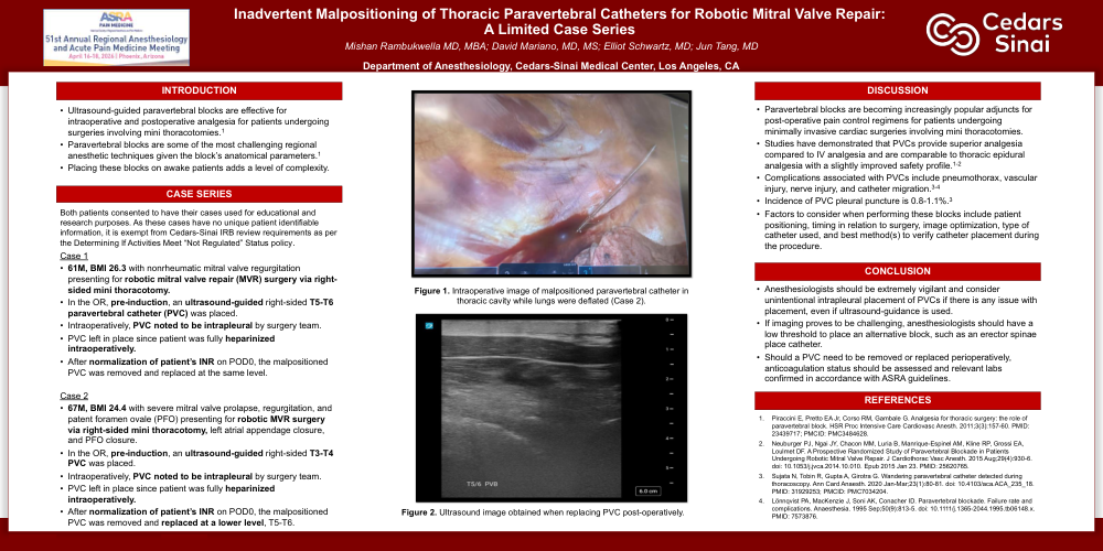
A 67M, BMI 24.4, with severe mitral valve prolapse, regurgitation, and patent foramen ovale (PFO) presented for robotic MVR surgery via right-sided mini thoracotomy, left atrial appendage closure, and PFO closure. Before induction of general anesthesia with ASA standard monitors, an ultrasound-guided T3-T4 PVC was placed. Intraoperatively, the PVC tip was noted to be intrapleural in the right thoracic cavity. The decision was made to leave the catheter in place until the patient’s coagulation status normalized. After the INR normalized on POD0, the malpositioned, intrapleural catheter was removed, and a replacement catheter was placed at T6-T7 under ultrasound guidance. This catheter provided appropriate analgesia at the chest tube site prior to its removal without complications on POD4 after the patient’s chest tube was removed.
Discussion
Paravertebral blockade is a popular adjunct for post-operative pain regimens in minimally invasive cardiac surgeries. Studies demonstrated that paravertebral blockade provides superior analgesia to intravenous analgesia and is comparable to thoracic epidural analgesia [1,2]. However, these blocks' risks include pleural puncture (0.8 - 1.1%), pneumothorax, vascular injury, nerve injury, and catheter migration [3,4]. Anesthesiologists must remain vigilant with these blocks and should consider alternative blocks if there is concern for malposition during placement.
References
[1] Piraccini E, Pretto EA Jr, Corso RM, Gambale G. Analgesia for thoracic surgery: the role of paravertebral block. HSR Proc Intensive Care Cardiovasc Anesth. 2011;3(3):157-60. PMID: 23439717; PMCID: PMC3484628.
[2] Neuburger PJ, Ngai JY, Chacon MM, Luria B, Manrique-Espinel AM, Kline RP, Grossi EA, Loulmet DF. A Prospective Randomized Study of Paravertebral Blockade in Patients Undergoing Robotic Mitral Valve Repair. J Cardiothorac Vasc Anesth. 2015 Aug;29(4):930-6. doi: 10.1053/j.jvca.2014.10.010. Epub 2015 Jan 23. PMID: 25620765.
[3] Sujata N, Tobin R, Gupta A, Girotra G. Wandering paravertebral catheter detected during thoracoscopy. Ann Card Anaesth. 2020 Jan-Mar;23(1):80-81. doi: 10.4103/aca.ACA_235_18. PMID: 31929253; PMCID: PMC7034204.
[4] Lönnqvist PA, MacKenzie J, Soni AK, Conacher ID. Paravertebral blockade. Failure rate and complications. Anaesthesia. 1995 Sep;50(9):813-5. doi: 10.1111/j.1365-2044.1995.tb06148.x. PMID: 7573876.
Figure 1. Intraoperative image of malpositioned paravertebral catheter in thoracic cavity while lungs were deflated (Case 2).