395 posters, 1 audios, 13 topics, 29 sessions, 1,056 authors, 461 institutions
ePostersLive by SciGen Technologies S.A. All rights reserved.
51st Annual Regional Anesthesiology and Acute Pain Medicine Meeting
April 16 - 18, 2026 | Phoenix, Arizona

2316295
Secondary failure of thoracic epidural after fluoroscopic confirmation and possible solutions to these barriers
Poster Presenter
Authors
Affiliations
Part of Topic
Medically Challenging Cases
Background
Thoracic epidural placement is considered the gold standard for the treatment of postoperative pain for major abdominal surgeries. The primary success of thoracic epidural placement is greatly enhanced with fluoroscopically guided techniques; however, this does not eliminate secondary failure due to epidural anatomy (1,2).
Epidural ligaments, septations, or prior trauma can influence the appropriate distribution of epidural local anesthetics. The following case describes the secondary failure of a fluoroscopically guided epidural.
As this case report is devoid of patient identifiable information, it is exempt from IRB review requirements as per Dartmouth-Health policy. Patient-informed consent was obtained for the use of the de-identified case information and images presented.
Objectives
- Describe the placement of fluoroscopically guided epidurals for thoracic epidural analgesia.
- Describe a case of secondary epidural failure despite fluoroscopic confirmation.
- Suggest practical strategies that may overcome coverage deficits from secondary failure.
Methods
Fluoroscopic epidural placement is completed by first obtaining a posterior–anterior (PA) image, with a slight caudad tilt (~7.5 degrees), of the prone patient to locate midline and vertebral levels. The epidural needle is advanced with periodic PA images to guide needle position over the intended level of insertion until tactile engagement in ligament is appreciated.
Loss of resistance is found using standard technique with a saline-filled syringe. Once the epidural space is entered, the epidural catheter is advanced with periodic PA imaging to verify catheter trajectory and tip location to the desired level based on the surgical procedure.
An iodinated contrast (2 mL) is injected through the catheter, followed by PA and lateral images, to confirm a positive epidurogram.
Case Description
A 52-year-old male with no significant past medical history was admitted for a gastric/duodenal obstruction secondary to a pancreatic head mass and scheduled for a Whipple procedure with an epidural for postoperative pain control.
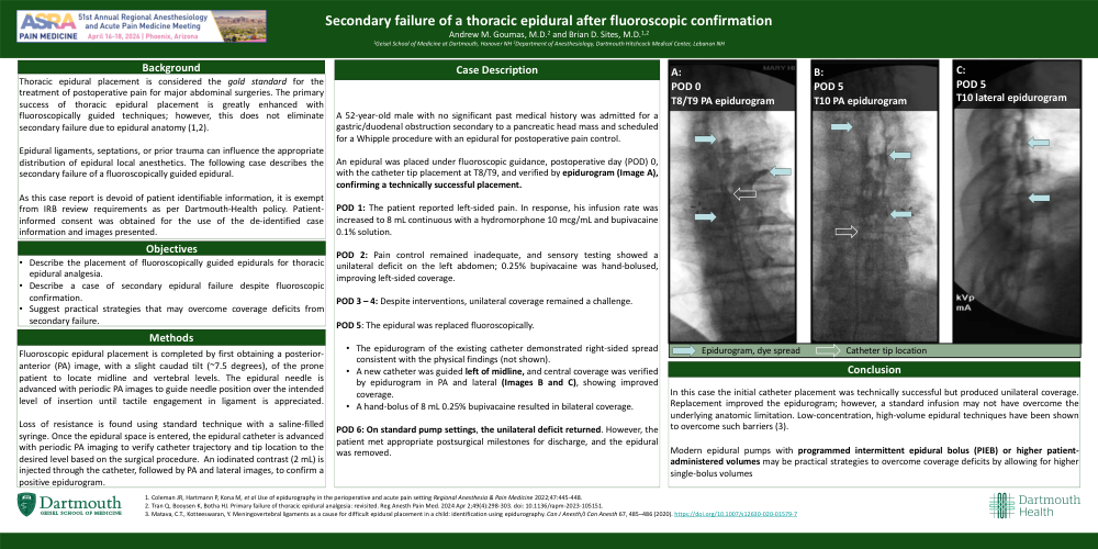
An epidural was placed under fluoroscopic guidance on postoperative day (POD) 0, with catheter tip placement at T8/T9 and verified by epidurogram (Image A), confirming a technically successful placement.
POD 1:
The patient reported left-sided pain. The infusion rate was increased to 8 mL continuous with hydromorphone 10 mcg/mL and bupivacaine 0.1%.
POD 2:
Pain control remained inadequate. Sensory testing showed a unilateral deficit on the left abdomen. A hand bolus of 0.25% bupivacaine improved left-sided coverage.
POD 3–4:
Despite interventions, unilateral coverage remained a challenge.
POD 5:
The epidural was replaced fluoroscopically. The epidurogram of the existing catheter demonstrated right-sided spread consistent with the physical findings (image not shown).
A new catheter was guided left of midline, and central coverage was verified by epidurogram in PA and lateral views (Images B and C), showing improved coverage. A hand bolus of 8 mL 0.25% bupivacaine resulted in bilateral coverage.
POD 6:
On standard pump settings, the unilateral deficit returned. The patient met appropriate postsurgical milestones for discharge, and the epidural was removed.
Conclusion
In this case, the initial catheter placement was technically successful but produced unilateral coverage. Replacement improved the epidurogram; however, a standard infusion may not have overcome the underlying anatomic limitation.
Low-concentration, high-volume epidural techniques have been shown to overcome such barriers (3). Modern epidural pumps with programmed intermittent epidural bolus (PIEB) or higher patient-administered volumes may be practical strategies to overcome coverage deficits by allowing for higher single-bolus volumes.